Mammary gland affection quantification image evaluation system and using method thereof

An evaluation system and imaging technology, applied in the field of medical diagnostic equipment for breast disease, can solve the problem of lack of quantitative evaluation indicators for imaging diagnosis

Active Publication Date: 2008-08-06
李立
View PDF0 Cites 24 Cited by
  • Summary
  • Abstract
  • Description
  • Claims
  • Application Information

AI Technical Summary

Problems solved by technology

[0004] The purpose of the present invention is to overcome the problem of the lack of quantitative evaluation indicators in the existing mammogram image diagnosis, and to provide a quantitative image evaluation system for breast lesions, which can perform quantitative analysis on mammogram images , providing the predictive value of benign and malignant breast lesions, as well as the predictive value of tumor cell grade

Method used


Image

  • Mammary gland affection quantification image evaluation system and using method thereof

Examples


Embodiment 1

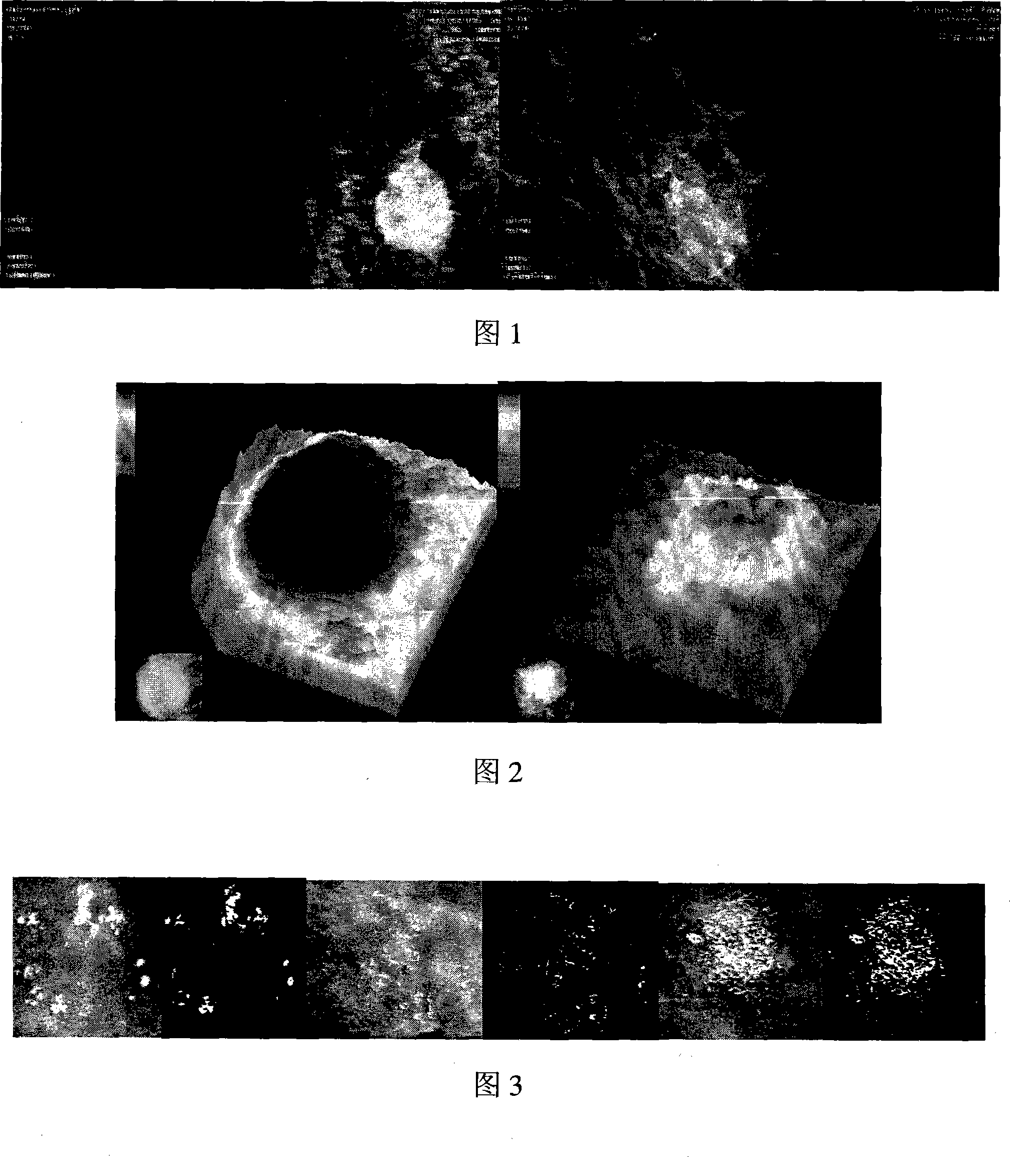
[0045] Image-Pro Plus software was used to automatically obtain the ROIs of benign and malignant tumors in mammography. The outline of the tumor boundary is shown in Figure 1; the two-dimensional and three-dimensional images of the tumor are shown in Figure 2. The growth and diffusion parameters of tumor cells in the region of interest were calculated using Image-Pro Plus software: the fractal dimension value was 1.14399; the internal heterogeneity value of the tumor was 0.02818. Introduce clinical parameters: age 46 years; history of breast disease (no); history of breastfeeding (yes). Using the linear regression equation: Y E =a*D F +b*H+c*C P +d*U+e*V+f*W, through calculation, the quantitative parameters of tumor cell growth and diffusion and clinical parameters are weighted differently, and the predicted value of benign and malignant tumors is Y E =0, benign lesion. Clinicopathological findings: benign breast fibroadenoma. The imaging evaluation results were consisten...

Embodiment 2

[0047] Use the Image-Pro Plus software to automatically obtain the region of interest of the mammogram, the processed graphics are similar to Fig. 1 and Fig. 2 described in Example 1, use the Image-Pro Plus software to calculate the tumor cell growth in the region of interest Diffusion parameters: the value of fractal dimension was 1.17089; the value of intra-tumor heterogeneity was 0.1783. Introduce clinical parameters: age 45 years old; history of breast disease (no); history of breastfeeding (yes). Using the linear regression equation: Y E =a*D F +b*H+c*C P +d*U+e*V+f*W, through calculation, the quantitative parameters of tumor cell growth and diffusion and clinical parameters are weighted differently, and the predicted value of benign and malignant tumors is Y E =1, it is a malignant lesion (breast cancer), and the grade of malignant tumor cells is close to grade I. Clinicopathological results: breast cancer, pathological grade I. The results of imaging evaluation wer...

Embodiment 3

[0049] Use Image-Pro Plus software to automatically obtain the interest area of ​​mammography, and use Image-Pro Plus software to calculate the growth and diffusion parameters of tumor cells in the area of ​​interest: the fractal dimension value is 1.19336; the tumor internal heterogeneity value is 0.53494 . The introduction of clinical parameters: age 50 years old; breast disease history (yes); lactation history (yes). Using the linear regression equation: Y E =a*D F +b*H+c*C P +d*U+e*V+f*W, through calculation, the quantitative parameters of tumor cell growth and diffusion and clinical parameters are weighted differently, and the predicted value of benign and malignant tumors is Y E =1.98, it is a malignant lesion (breast cancer), and the grade of malignant tumor cells is close to grade II. Clinicopathological results: breast cancer, pathological grade II. The imaging evaluation results were consistent with the pathological results, and were very similar to the patholog...


PUM


Abstract

The invention provides a quantitative image evaluation system of breast lesions and an application method thereof. The quantitative image evaluation system adopts fractal technology and graphic analysis in tumor medical image analysis and tumor hazard rate evaluation, establishes and adopts a nonlinear data model for growth and diffusion of breast lesion cells. The nonlinear data model comprises morphological characteristic parameters of growth, diffusion and calcification of mammary tumor cells and clinical parameters as well. The quantitative image evaluation system of breast lesions of the invention adopts the nonlinear data model for growth and diffusion of breast lesion cells and comprises the morphological characteristic parameters of growth, diffusion and calcification of mammary tumor cells and the clinical parameters as well, calculates predictive values of benign and malignant breast lesions by mammography and predictive values of tumor cell classification; therefore, the quantitative image evaluation system can be widely used in mammography diagnosis and mammography screening.

Description

technical field [0001] The invention belongs to a medical diagnostic device for breast disease, and in particular relates to a quantitative image evaluation system applied to mammograms and the application of the system. technical background [0002] Breast imaging examinations, especially mammography, provide an important basis for the diagnosis, staging, and efficacy evaluation of breast tumors, and have been used in breast screening. Unfortunately, in the current clinical diagnosis, conventional imaging diagnosis is limited to the measurement of tumor size and simple shape factor and some qualitative evaluations, and there is a lack of quantitative imaging evaluation indicators in clinical applications. With the continuous improvement of medical image quantitative analysis technology, it is also necessary to evaluate the growth and diffusion characteristics of different tumor types through image quantitative analysis in the basic research of tumors. I...

Claims


Application Information

Patent Timeline
Patent Type & Authority Applications(China)
IPC IPC(8): A61B6/02G06F19/00
Inventor 李立邵元智刘立志别梦杰李婵婵钟伟荣崔春艳陈第虎
Owner 李立
Why Eureka
Try Eureka