Disposable uterine cavity and vagina visual combination probe

A combined probe and one-time technology, applied in colposcopy, medical science, endoscopy, etc., can solve the problems of surgical instruments that are too thick, delay treatment, increase the risk of cross-infection, etc., achieve intuitive imaging effects and reduce pain Effect

Inactive Publication Date: 2009-03-18
上海家宝医学保健科技有限公司
View PDF0 Cites 1 Cited by
  • Summary
  • Abstract
  • Description
  • Claims
  • Application Information

AI Technical Summary

Problems solved by technology

At present, in routine gynecological examinations and operations, B-ultrasound, laparoscopy or other imaging systems are often used to observe the pre- and post-operative status and effects, but these devices have low image resolution and too thick surgical instruments. (generally between 10 and 26mm), and it is not a one-time use problem
Therefore, it is difficult to visually determine the location of the lesion. At the same time, in the detection of some uterine items, it is necessary to perform uterine expansion or uterine expansion, which will inevitably cause accidental damage sometimes.
In addition, due to the phenomenon of cross-use of equipment, it is difficult to avoid the possibility of cross-infection
[0003] It is for the above reasons, coupled with the special structure of the uterus, that it is impossible to implement direct imaging and imaging techniques for positioning; only by using other more complicated auxiliary operation methods can the ideal detection in the uterus be completed.
As a result, some gynecological lesions are often not discovered in time, thereby delaying the best time for treatment, endangering women's physical and mental health, and cross-infection of surgical instruments will virtually increase the risk of cross-infection in the female population

Method used


Image

  • Disposable uterine cavity and vagina visual combination probe
  • Disposable uterine cavity and vagina visual combination probe
  • Disposable uterine cavity and vagina visual combination probe

Examples


Embodiment Construction

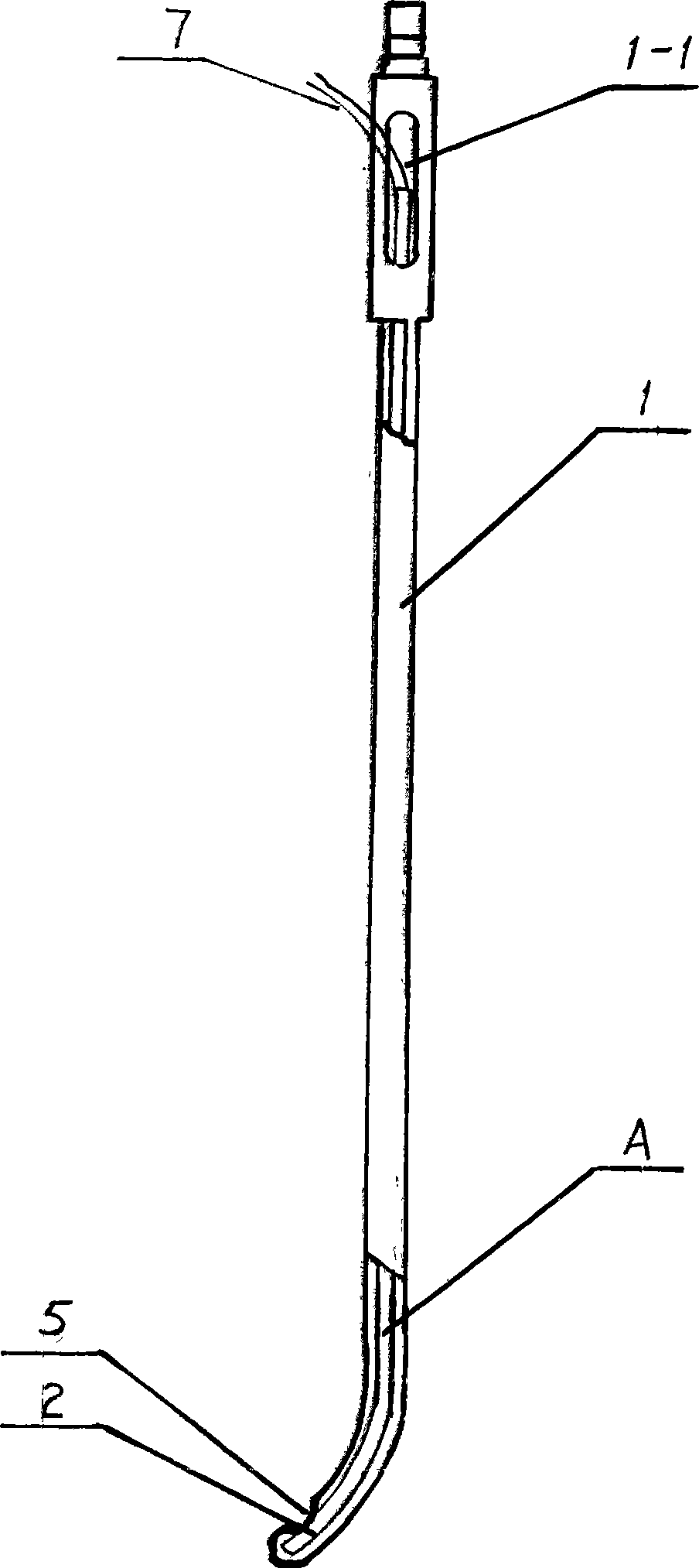
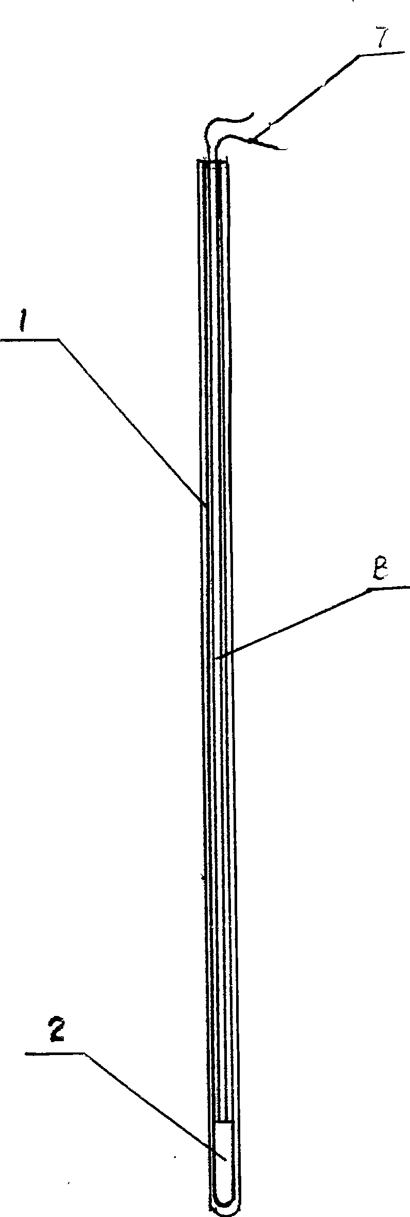
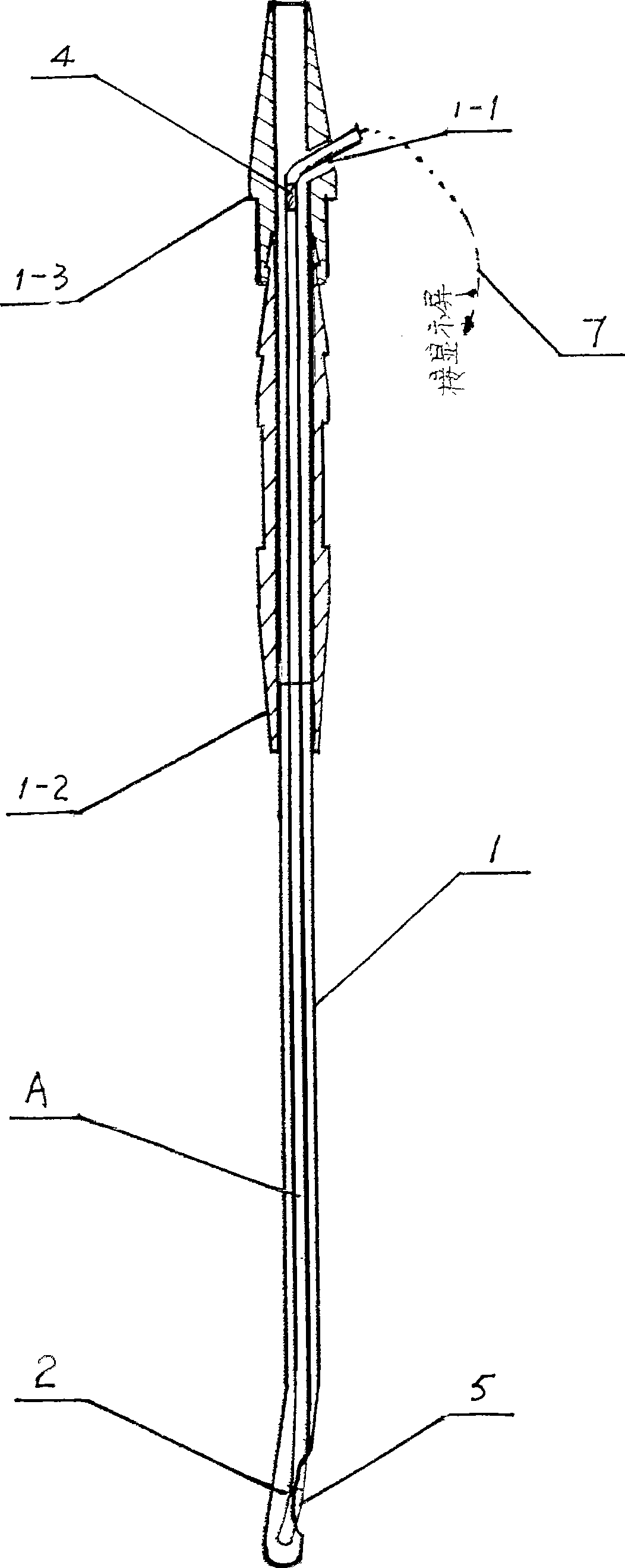
[0021] like figure 1 This disposable uterine cavity and vagina visible combined probe (embodiment 1) as shown, it comprises a sleeve tube 1 that can stretch into the uterine cavity, and is located in the sleeve tube hollow cavity, can extract out and replace at will. And a visible tubular probe body A composed of a miniature camera 2, a flexible printed board 3 and a signal output terminal 4; and connected to the imaging system through the lead-out line 7 of the signal output terminal.

[0022] The micro-camera 2 of the visible probe A has a diameter of 1 / 18-1 / 10 inch.

[0023] The end of the flexible sleeve 4 is provided with an oval aperture 5 matching the miniature camera 2

[0024] The miniature camera 2 at the front end of the camera A should be placed within the range of 3-8 mm at the front end of the flexible transparent sleeve 4, generally 5 mm.

[0025] In this disposable uterine cavity and vaginal visible combined probe, the practical flexible sleeve is actually a ...


PUM


Abstract

The invention relates to a one-off visual combination probe for uterine cavities and vaginas. The invention is characterized in that: the combination probe comprises a flexible bushing which can extend to a uterine cavity and a tube-shaped visual probe body A which is arranged in the hollow cavity of the flexible bushing, can be drawn out freely and comprises a micro camera, a flexible printed plate and signal output end; and the probe body A is led out by a lead which is connected with a display equipment from the rear part. As micro visual camera technology is applied in monitoring and imaging of uterine cavities and vaginas, the affected part of pathological changes of uterine cavities and vaginas of patients can be displayed on the screen clearly and reliably, one-off check of uterine cavities and vaginas without hurt or pain is realized, the pain of the patients is alleviated and the possibility of cross infection is avoided; meanwhile, intuitionistic imaging effect without operation is provided for diagnosis of doctors.

Description

technical field [0001] The invention relates to a gynecological organ and tissue detection instrument, in particular to a disposable uterine cavity and vaginal visible combined probe for detection of uterine cavity and vaginal lesions. Background technique [0002] The uterus is not only an important reproductive organ of women, but also a site that is prone to gynecological lesions in the female population. Uterine erosion, uterine cancer, and cervical cancer have become the main killers that endanger women's physical and mental health; at the same time, anti-early pregnancy has become an important aspect of family planning work. Since the early detection of the above-mentioned lesions and physiological conditions has a higher success rate for curing the above-mentioned gynecological diseases, the effect of treating early pregnancy is also the best. At present, in routine gynecological examinations and operations, B-ultrasound, laparoscopy or other imaging systems are ofte...

Claims


Application Information

Patent Timeline
Patent Type & Authority Applications(China)
IPC IPC(8): A61B1/303A61B1/04
Inventor 徐明
Owner 上海家宝医学保健科技有限公司
Why Eureka
Try Eureka