Device for superposed magnetic resonance and positron emission tomography imaging

A technology of positron emission and magnetic resonance tomography, which is applied in the direction of radiodiagnostic instruments, measuring devices, computer tomography scanners, etc.

Inactive Publication Date: 2009-06-10
SIEMENS AG
View PDF0 Cites 12 Cited by
  • Summary
  • Abstract
  • Description
  • Claims
  • Application Information

AI Technical Summary

Problems solved by technology

In such a construction, the problem arises that eddy currents are excited by gradient fields in the shielding of the PET detector, leading to vibrations and heating

Method used


Image

  • Device for superposed magnetic resonance and positron emission tomography imaging
  • Device for superposed magnetic resonance and positron emission tomography imaging
  • Device for superposed magnetic resonance and positron emission tomography imaging

Examples


Embodiment Construction

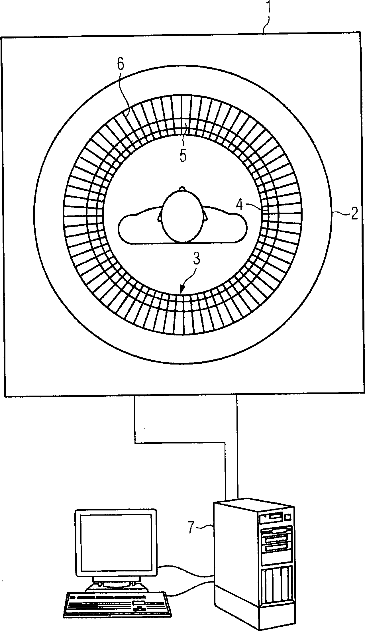
[0017] Embodiments of the present invention are preferably used on combined MR-PET devices. The combined device has the advantage that MR data and PET data can be acquired concentrically. This makes it possible to precisely define an examination volume within a region of interest using data from a first modality (PET) and to use this information in another modality (eg magnetic resonance). Although it is possible to transmit the volume information of the region of interest from an external PET system to the MR system, this increases the outlay for registering the data. In general, all data that can be determined using magnetic resonance or other imaging methods can be determined on selected regions of interest in the PET data set. For example, instead of spectroscopic data, fMRI data, diffusion maps, T1- or T2-weighted images or quantitative parametric maps can also be acquired in the region of interest by means of magnetic resonance examinations. Likewise, computed tomograp...


PUM


Abstract

The present invention provides a device for superposed magnetic resonance and positron emission tomography imaging. The device includes a gradient coil and a positron emission tomography unit (PET unit). The PET unit is arranged within the gradient coil and has a first shield against radiofrequency radiation which in part surrounds the PET unit, and a second shield against radiofrequency radiation is arranged on the gradient coil. The first shield is connected to the second shield to form a shield which is at least partly closed. This makes a closed shield for the PET unit possible, which nevertheless is still easily accessible for maintenance purposes due to the two-part design of the shield.

Description

technical field [0001] The invention relates to a device for superimposed magnetic resonance tomography and positron emission tomography. Background technique [0002] In recent years, in addition to magnetic resonance tomography (MR), positron emission tomography (PET) has been increasingly used in medical diagnosis. MR is an imaging method used to visualize structures and cross-sections inside the body, while PET can visualize and quantify metabolic activity in the body. [0003] PET exploits the specific characteristics of positron radiation sources and positron annihilation in order to quantitatively determine the function of organs or cellular regions. Here, a radiopharmaceutical labeled with a radionuclide is administered to the patient prior to the examination. In the case of radionuclide decay, a positron is released which, after a short distance, interacts with an electron, resulting in the so-called annihilation. Here, two gamma quanta are produced, which fly ou...

Claims


Application Information

Patent Timeline
Patent Type & Authority Applications(China)
IPC IPC(8): A61B5/055A61B6/03A61B6/06
CPCA61B6/037G01R33/422G01R33/481A61B6/4417
Inventor 沃尔夫冈·伦兹斯蒂芬·斯托克
Owner SIEMENS AG
Why Eureka
Try Eureka