Device for measuring biology tissue inner light depth
A biological tissue and depth technology, applied in diagnostic recording/measurement, medical science, sensors, etc., can solve problems such as real-time tracking and positioning application limitations, tissue heterogeneity, light scattering, etc.
Active Publication Date: 2010-09-08
SUZHOU ZHONGKE ADVANCED TECH RES INST CO LTD
View PDF0 Cites 0 Cited by
- Summary
- Abstract
- Description
- Claims
- Application Information
AI Technical Summary
Problems solved by technology
At present, researchers are also trying to implement real-time image guidance with commonly used ultrasound imaging, magnetic resonance imaging, computed tomography imaging, fluorescence imaging and bioluminescence imaging, but they all encounter some difficulties
Most of the existing imaging methods such as ultrasound, nuclear magnetic resonance, and computed tomography are difficult to use in clinical operations due to issues such as tissue differentiation, real-time imaging, or radiation safety; fluorescence imaging technology is affected by imaging depth, signal-to-noise ratio, etc. It has great limitations in actual surgery. Due to the influence of the angle of the light source, it is also greatly restricted in the application of real-time tracking and positioning of moving organs.
Bioluminescent imaging mainly includes surface imaging and tomographic imaging, but it is difficult to take out clear images from tissues due to tissue inhomogeneity and light scattering.
Bioluminescence imaging can be divided into BLI (Bioluminescent Imaging) and BLT (Bioluminescent Tomography) according to different imaging methods, but BLI outputs a two-dimensional image, which mainly detects the optical signal of the biological surface, and cannot measure the depth of the luminescent light source in the living organism. ; and BLT due to the limitations of bioluminescent imaging itself, the reconstruction accuracy is far from meeting the requirements
Generally speaking, none of the above-mentioned imaging technologies can accurately give the depth information of the tumor in the living body during the actual operation, and there is no small and cheap tumor depth measurement method and measurement device suitable for real-time navigation in clinical operations
Method used
View moreImage
Smart ImageViewing Examples


Examples
Embodiment Construction
Login to View More PUM
 Abstract
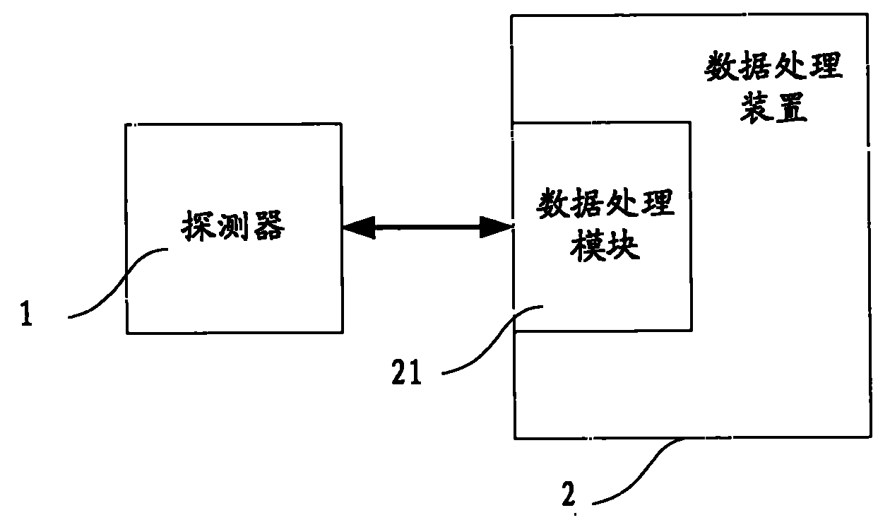
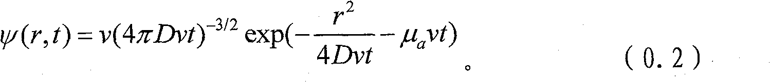
The invention relates to a method for measuring light source depth in biological tissue, the light source comprises a part of abnormal structure capable of sending continuous light waves in the biological tissue, the method comprises following steps: measuring the light strength signals of the light sensor positions by at least three light sensors arranged on a detector respectively; calculating the depth of a luminous part in the tissue according to a distance between the obtained light strength signal and the light sensor corresponding to the light strength signal. The invention also discloses an apparatus using the measuring method. The invention has following beneficial effects: the depth information of the luminous light source in the tissue can be obtained, meanwhile, a smart and low-price tumor depth measuring apparatus of real capable of navigating in real time and suitable for clinical operation can be obtained.
Description
technical field The present invention relates to medical equipment, and more particularly, to a device for measuring the depth of a light source in biological tissue. Background technique Real-time image-guided technology can help doctors perform tumor resection surgery with a clearer view, which can greatly improve surgical accuracy and reduce surgical incisions. At present, researchers are also trying to implement real-time image guidance with commonly used ultrasound imaging, magnetic resonance imaging, computed tomography imaging, fluorescence imaging, and bioluminescence imaging, but they all encounter some difficulties. Most of the existing imaging methods such as ultrasound, nuclear magnetic resonance, and computed tomography are difficult to use in clinical operations due to issues such as tissue differentiation, real-time imaging, or radiation safety; fluorescence imaging technology is affected by imaging depth, signal-to-noise ratio, etc. It has great limitations ...
Claims
Login to View More Application Information
Patent Timeline
 Patent Type & Authority Patents(China)
IPC IPC(8): A61B5/107
Inventor 郑震欧石平
Owner SUZHOU ZHONGKE ADVANCED TECH RES INST CO LTD