3D model building method and system

A three-dimensional model and type of technology, applied in the medical field, can solve problems such as time-consuming and manpower-consuming, unsuitable for clinical individual research, low efficiency of three-dimensional models, etc., and achieve the effect of improving efficiency

Inactive Publication Date: 2009-08-05
SIEMENS SHANGHAI MEDICAL EQUIP LTD
View PDF0 Cites 28 Cited by
  • Summary
  • Abstract
  • Description
  • Claims
  • Application Information

AI Technical Summary

Problems solved by technology

In the prior art, the methods of manually measuring and reading specimens or specimen model slices and grinding slices are used to survey and map the shape coordinates of the fault layer by layer, and some directly measure the three-dimensional shape coordinates of specimens (such as laser mapping and surface rendering methods) to generate three-dimensional However, in these traditional methods, it is time-consuming and labor-intensive to use manual measurement and read

Method used


Image

  • 3D model building method and system
  • 3D model building method and system
  • 3D model building method and system

Examples


Embodiment Construction

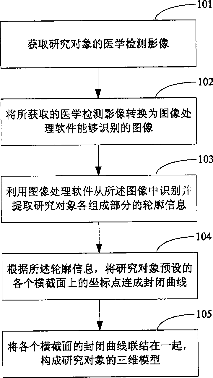
[0056]In the embodiment of the present invention, it is considered that imaging techniques such as computed tomography (CT) and magnetic resonance (Magnetic Resonance Imaging, MRI) are widely used in clinical diagnosis, and imaging techniques such as CT and MRI have many advantages For example, for CT imaging: CT tomographic image deformation is small, the information obtained is comprehensive and accurate, and can reflect more detailed and complex structures; scanning is non-invasive and destructive, and the integrity of the examined object is preserved; each tomographic two Dimensional information is positioned accurately, and the spatial positions are arranged in sequence; high resolution, easy for image segmentation; reusable, etc. For MRI imaging: MRI imaging has no bony artifacts, and direct multi-directional (transverse, coronal, or any angle) slices can be made at will; higher soft tissue resolution capabilities, etc. For this reason, in the embodiment of the present i...


PUM


Abstract

The invention discloses a method for establishing a three-dimensional model. The method includes the steps: acquiring the medical detection image of a research object; converting the medical detection image to an image that can be identified by image processing software; utilizing the image processing software to identify and extract the profile information of all the components of the research object from the image; connecting the coordinate points on each cross section preset by the research object into a closed curve according to the profile information; and connecting the closed curves of all the cross sections together to form the three-dimensional model of the research object. Besides, the invention also discloses a system for establishing the three-dimensional model. When the technical proposal disclosed in the invention is used for establishing the three-dimensional model, manual surveying and mapping are not needed, thus improving the efficiency of establishing the three-dimensional model. In addition, as the medical detection image of a clinical individual can be acquired any time, the invention is not necessarily limited to a specified specimen and consequently applicable to clinical individual research.

Description

technical field [0001] The invention relates to the field of medicine, in particular to a method and system for establishing a three-dimensional model. Background technique [0002] With the rapid development of computer image processing technology, it is becoming more and more popular to use image processing software for research or auxiliary treatment in the medical field. For example, use image processing software to analyze the force of the implant, analyze the impact of different shapes (or materials) of the implant on the implant matrix; simulate the operation on the three-dimensional model, and calculate various parameters of the operation (such as the position, direction and size of the operation). ), predict postoperative results; carry out various medical statistical activities, etc. [0003] But no matter what kind of research or adjuvant therapy work, it is necessary to establish a three-dimensional model of the research object before using image processing soft...

Claims


Application Information

Patent Timeline
IPC IPC(8): G06T17/00A61B5/055A61B6/02
Inventor 曹斌
Owner SIEMENS SHANGHAI MEDICAL EQUIP LTD
Why Eureka
Try Eureka