Method for automatically separating adherent hyaline-vascular type lung nodule in CT image

A CT image, automatic segmentation technology, applied in image analysis, image data processing, instruments, etc., can solve the problems of speed and accuracy can not meet the application requirements, time-consuming and so on

Active Publication Date: 2009-08-26
NEUSOFT MEDICAL SYST CO LTD
View PDF0 Cites 36 Cited by
  • Summary
  • Abstract
  • Description
  • Claims
  • Application Information

AI Technical Summary

Problems solved by technology

[0005] In view of the problems that the nodule segmentation method adopted in the prior art is time-consuming, and the speed and accuracy cannot meet the application requirements, the technical problem to be sol

Method used


Image

  • Method for automatically separating adherent hyaline-vascular type lung nodule in CT image
  • Method for automatically separating adherent hyaline-vascular type lung nodule in CT image
  • Method for automatically separating adherent hyaline-vascular type lung nodule in CT image

Examples


Embodiment 1

[0073] In this embodiment, the automatic segmentation of vascular pulmonary nodules in two-dimensional CT images is taken as an example.

[0074] The present invention is an automatic segmentation method for adhesive vascular pulmonary nodules in CT images, which can quickly and adaptively obtain Mean-shift (mean value shift) bandwidth parameters, and is used for automatic segmentation of adhesive vascular pulmonary nodules in CT images. In this way, the speed and accuracy requirements of the vascular adhesion type pulmonary nodule segmentation algorithm are met at the same time. Such as Figure 4 Shown, the concrete steps of the inventive method are as follows:

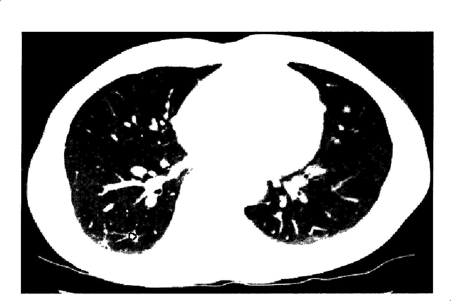
[0075] (1) Input the region of interest of the CT image containing the adherent vascular pulmonary nodules;

[0076] (2) Preprocessing is performed on the above-mentioned region of interest to obtain the foreground area of ​​the region of interest;

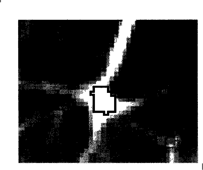
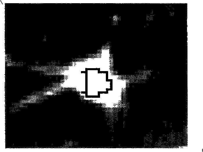
[0077] (3) Extract the flow direction feature based on the relat...

Embodiment 2

[0141] In this embodiment, the automatic segmentation of vascular pulmonary nodules in three-dimensional CT images is taken as an example.

[0142] The concrete steps of the inventive method are as follows:

[0143] (1) Input the body of interest containing the CT image of the adherent vascular pulmonary nodule;

[0144] (2) Preprocessing the above-mentioned body of interest to obtain the foreground area of ​​the body of interest, and project the foreground area onto a two-dimensional plane using a maximum density projection method;

[0145] (3) Extract the flow direction feature based on the relationship matrix in the foreground area projection on the above-mentioned two-dimensional plane;

[0146] (4) Establishing an adhesion vessel type pulmonary nodule model based on the above-mentioned flow direction characteristic direction angle;

[0147] (5) Estimating model parameters based on the expected maximum method in the above model;

[0148] (6) Utilize above-mentioned mode...


PUM


Abstract

The invention relates to a method for automatically separating adherent hyaline-vascular type lung nodule in a CT image, comprising the following steps: (1) inputting an interested body of the CT image containing the adherent hyaline-vascular type lung nodule; (2) preprocessing the interested body, thus obtaining a front image area; (3) extracting flow direction characteristics based on a relational matrix on the front image area; (4) establishing an adherent hyaline-vascular type lung nodule model based on the flow direction characteristics, direction and angle; (5) estimating model parameters based on the expectation-maximization method in the model; (6) obtaining mean shift bandwidth parameters by calculating the parameters of the model; and (7) putting the mean shift bandwidth parameters into the mean shift clustering algorithm for automatic separation. The method of the invention can obtain the self-adapting bandwidth parameters in an accurate and rapid manner while applying the method for selecting the self-adapting bandwidth parameters to automatically separate adherent hyaline-vascular type lung nodule in a CT image, thus meeting the dual requirements on speed and accuracy in practical application.

Description

technical field [0001] The invention relates to an image processing method in the technical field of medicine, in particular to an automatic segmentation method for pulmonary nodules with adhesion vessels in CT images. Background technique [0002] Lung cancer has the highest mortality rate among all cancers. In clinical practice, the diagnosis of benign and malignant lung cancer is of great significance. Pulmonary nodules are imaging manifestations of lung cancer. The growth rate of pulmonary nodules is an indicator for distinguishing benign from malignant. Computer aided detection (CAD) system provides a new method for the quantitative diagnosis of benign and malignant pulmonary nodules. It should automatically help physicians segment a nodule detected on an image in order to measure its volume and calculate its doubling rate over time. The doubling ratio refers to the size of nodules in two images of the same patient compared at different times. If the nodule volume i...

Claims


Application Information

Patent Timeline
IPC IPC(8): G06T7/00G06K9/34A61B6/03
Inventor 康雁孙申申赵宏
Owner NEUSOFT MEDICAL SYST CO LTD
Why Eureka
Try Eureka