Method for freeze preservation of tissue-derived cell
A cryopreservation and tissue technology, which is applied to the preservation of human or animal bodies, tissue culture, animal cells, etc., can solve the problems of labor and time, and achieve the effect of low-cost and high-efficiency acquisition
- Summary
- Abstract
- Description
- Claims
- Application Information
AI Technical Summary
Problems solved by technology
Method used
Image

Examples
Embodiment 1
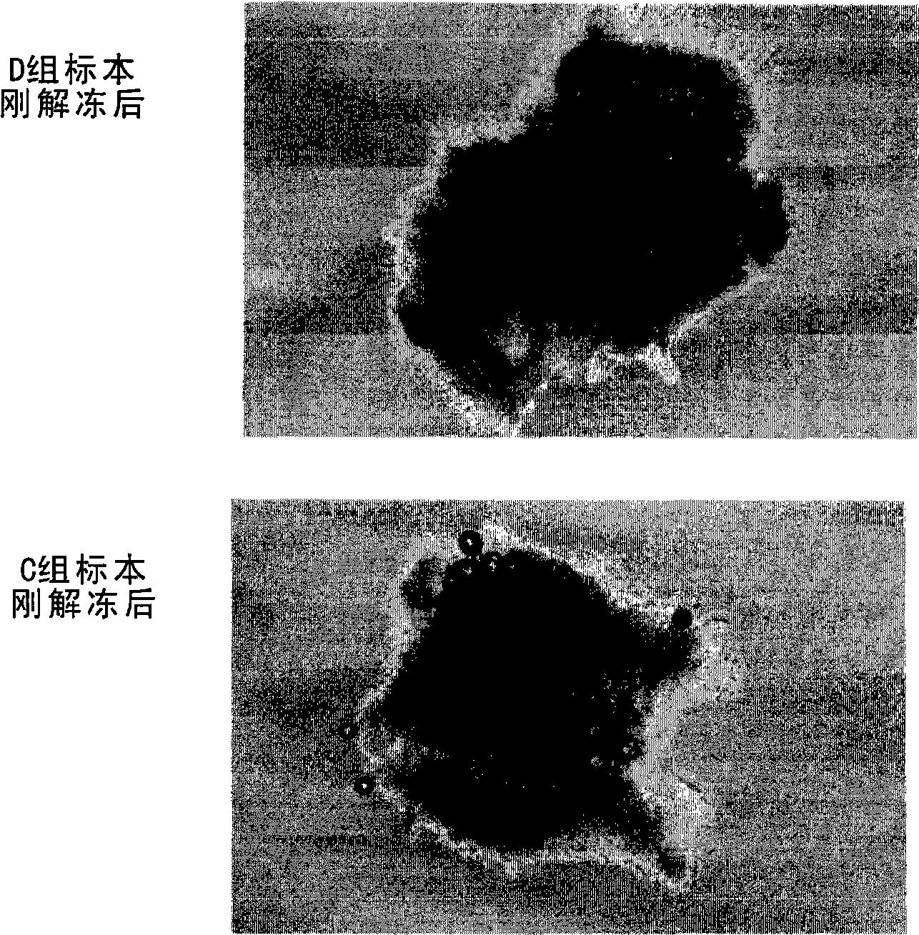
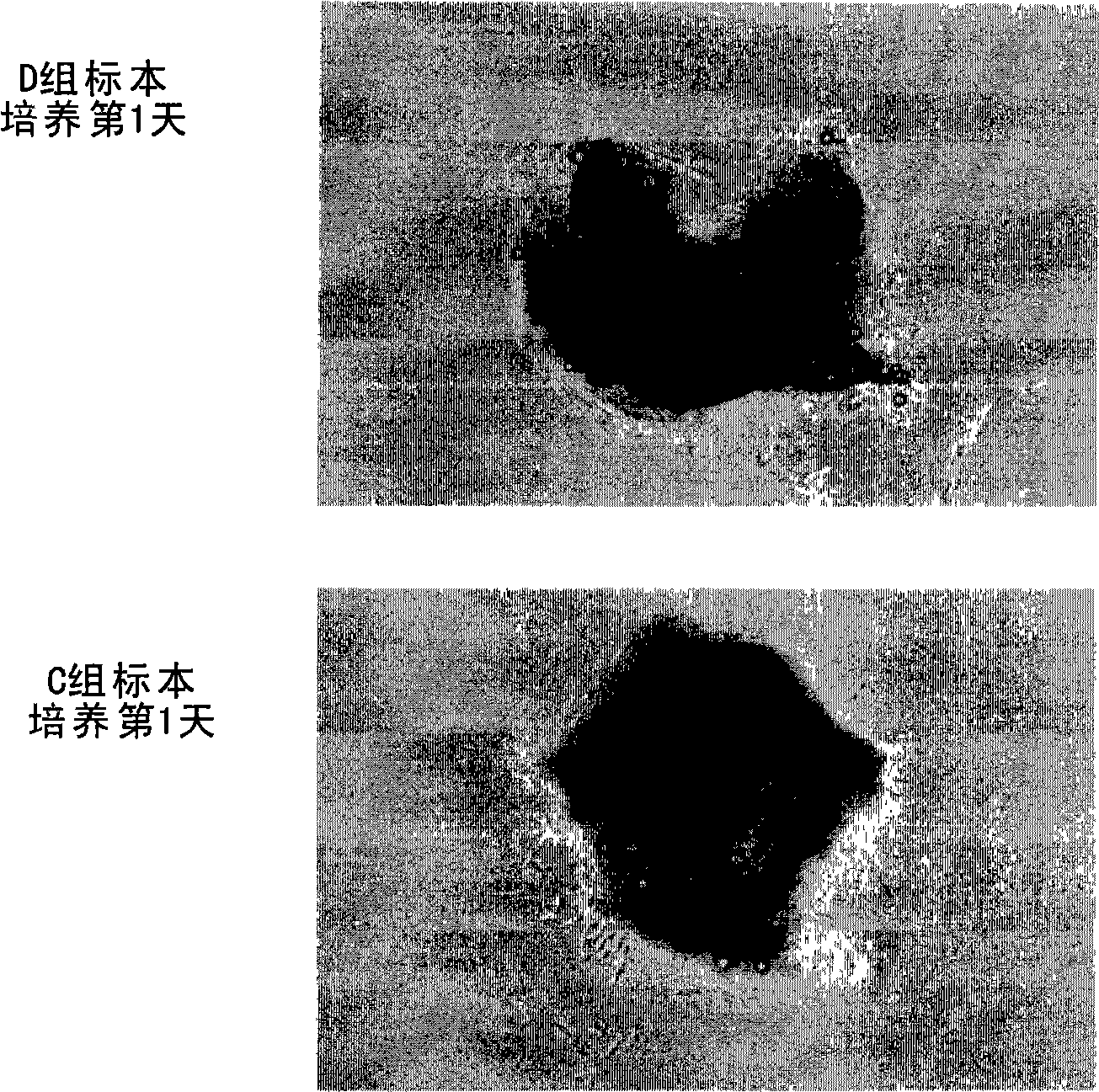
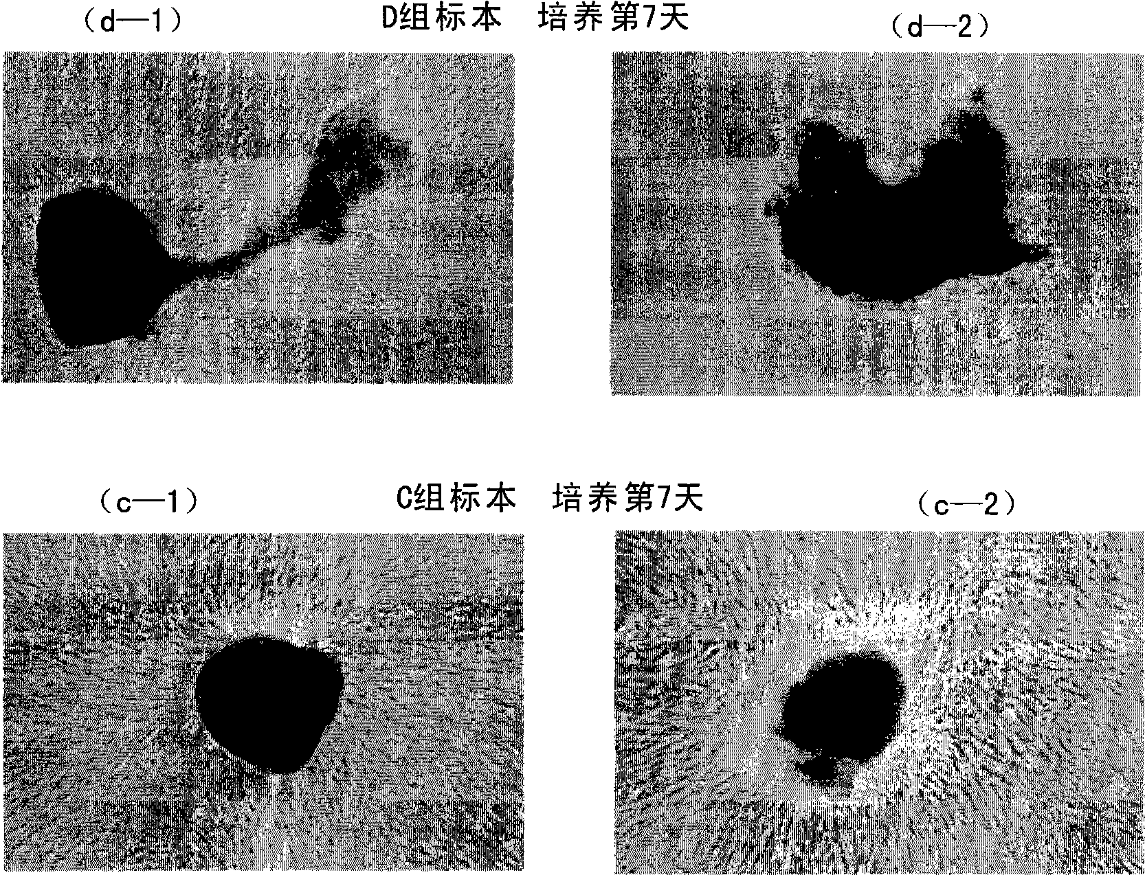
[0039] Three adult females (ages 33, 27, and 35 years old) and one adult male (age 50 years old) obtained their skins and performed the following experiments with their own consent. After each subject was anesthetized, a full-thickness skin of 1 mm to 3 mm was extracted from the back of the auricle. Immediately perform 2-hour enzyme (dispase2000 unit / ml, contract alcohol) treatment, separate the full-thickness skin into epidermis and dermis, discard the epidermis, and finely cut the dermis (about 0.5mm in diameter) to make 10 sliced specimens. The obtained 10 specimen slices were divided into 2 groups for the following experiments.
[0040] Group D (direct group): The specimen sections were immersed in cryopreservation solution (10% DMSO, 80% αMEM, 10% human serum) or commercially available cell cryopreservation solution Cellbanker (registered trademark) 1 (Shici) without any treatment. Field Co., Ltd.), stored at -80°C for 12 hours, and then frozen in liquid nitrogen for 2...
PUM
 Abstract
Description
Claims
Application Information
PatSnap Eureka turns technology decisions into work you can execute. Powered by our Innovation Knowledge Graph, it runs expert workflows across engineering, life sciences, materials and intellectual property. Get your review-ready output in minutes.


