
Method for extracting respiratory movement parameter from one-arm X-ray radiography picture

A technology of respiratory movement and shadow image, which is applied in image data processing, equipment for radiological diagnosis, image analysis, etc.

Active Publication Date: 2010-07-14
HUAZHONG UNIV OF SCI & TECH
View PDF0 Cites 25 Cited by
  • Summary
  • Abstract
  • Description
  • Claims
  • Application Information

AI Technical Summary

Problems solved by technology

Relatively speaking, this method can obtain more reliable respiratory motion estimation results, but due to the constraints of dual-arm X-ray angiography conditions, it cannot be widely used in medical practice

Method used


Image

  • Method for extracting respiratory movement parameter from one-arm X-ray radiography picture
  • Method for extracting respiratory movement parameter from one-arm X-ray radiography picture
  • Method for extracting respiratory movement parameter from one-arm X-ray radiography picture

Examples


Embodiment Construction

[0043] The present invention will be further described below in conjunction with accompanying drawing:

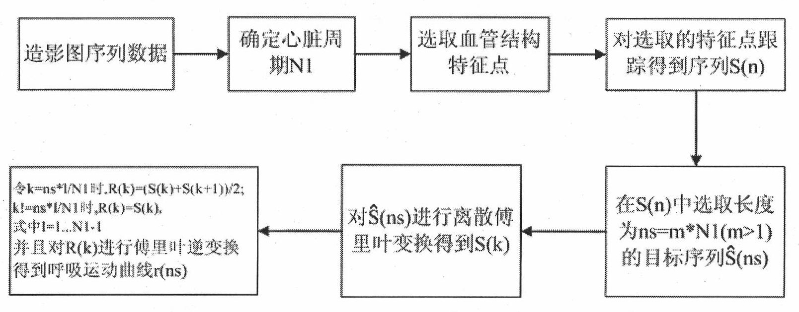
[0044] The present invention proposes a method for extracting human respiratory motion parameters based on frequency domain filtering, such as figure 1 shown, including the following steps:

[0045] (1) Obtain a single-arm X-ray contrast image sequence of coronary vessels, and determine the cardiac motion cycle N1;

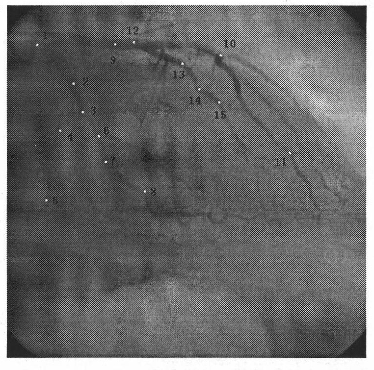
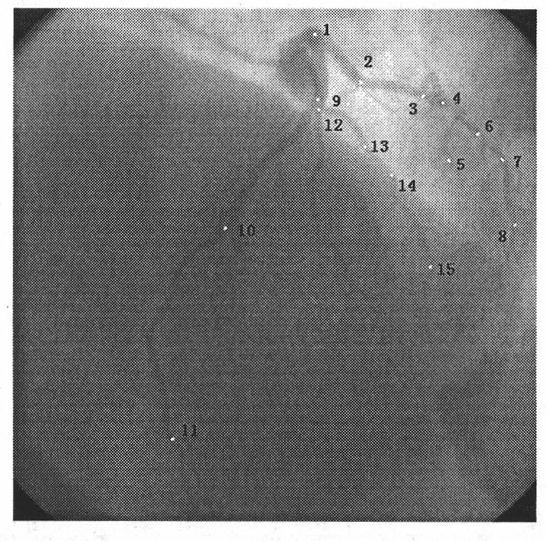
[0046] (2) Select the feature points of the vessel structure.

[0047] According to the guidance of physiological and anatomical knowledge, the feature points we mark need to be able to comprehensively reflect the movement information of the whole blood vessel. Therefore, the selected shape-specific points (ie structural feature points) generally include the starting point and ending point of each blood vessel segment. points, and each inflection point (the point with the greatest curvature) between the vessel segments. And in the sequence of contrast images...


PUM


Abstract

The invention relates to a method for extracting a respiratory movement parameter from a one-arm X-ray radiography picture, which belongs to the crossed field of digital signal processing and medical imaging, and aims at separating the respiratory movement from the complex movement of an actual human body heart. The method comprises the following steps: selecting a group of architectural feature points (including the head and tail points of a vessel section) on a coronal artery vessel and tracking the motion curve of the architectural feature point along with the time in a sequence; and separating to obtain a bi-dimensional respiratory movement curve by the Fourier expansion and frequency-domain filtering method. In addition, in the invention, in order to describe the respiratory movement more intuitively and effectively, the bi-dimensional respiratory movement obtained from two different radiography visual angles is rebuilt according to the three-dimensional reconstruction theory of the mid point of a radiography system, therefore the three-dimensional respiratory movement is obtained. The invention has the advantages of better extracting result of the respiratory movement and wider applicability and flexibility, and meets clinical requirements.

Description

technical field [0001] The invention belongs to the intersecting field of digital signal processing and medical imaging, and in particular relates to a method for extracting respiratory motion parameters from a single-arm X-ray contrast image. Background technique [0002] Rhythmic expansion and contraction of the chest to complete inhalation and exhalation, which is breathing movement. Breathing movement will cause the overall translational movement of the human heart in three-dimensional space. In the X-ray contrast system, due to the influence of respiratory movement, the coronary blood vessels will undergo two-dimensional translational movement on the imaging surface. [0003] Therefore, on the one hand, the coronary angiography image records the projection of the motion of the heart on the two-dimensional plane, and at the same time superimposes the two-dimensional translational movement of the coronary artery on the angiography plane caused by the respiratory movement...

Claims


Application Information

Patent Timeline
IPC IPC(8): A61B6/00G06T7/20G06T7/292
Inventor 张天序邓觐鹏孙祥平肖晶黎云曹治国桑农王国铸王芳
Owner HUAZHONG UNIV OF SCI & TECH
Learn More