Integral hard ultrasonic hysteroscope system

A technology of ultrasound system and hysteroscopy, applied in the direction of catheter, surgery, etc., can solve the problems of inconvenient hand operation, affecting image quality, instability, etc., and achieve the effect of improving operability, simple and convenient operation, and improving accuracy

Inactive Publication Date: 2010-07-28
广州市番禺区胆囊病研究所
View PDF8 Cites 23 Cited by
  • Summary
  • Abstract
  • Description
  • Claims
  • Application Information

AI Technical Summary

Problems solved by technology

[0002] At present, hysteroscopy and hysteroscopy technology have been widely used clinically, and are important tools for gynecological diagnosis and treatment. Changes in each layer of the cavity and surrounding organs, there is a hidden danger of insufficient diagnostic accuracy
The rigid ultrasonic hysteroscope system that has obtained the national utility model patent, its patent number is ZL200920006033.1 (announcement number: CN201299586Y), the method is to introduce micro-ultrasound technology, and the micro-ultrasound probe enters through the instrument channel of the hysteroscope Scan the uterus to obtain the conditions of the layers in the uterine cavity and between the uterine walls. The precise micro-ultrasound probe is easy to be damaged due to improper use, and due to the inconvenience and instability of manual operation, it is easy to cause ultrasound images and endoscopic images. The out-of-synchronization affects the quality of the image and directly reduces the accuracy of the doctor's diagnosis
[0003] There is currently no endoscopic tool that integrates rigid hysteroscopy with micro-ultrasound probe technology

Method used


Image

  • Integral hard ultrasonic hysteroscope system
  • Integral hard ultrasonic hysteroscope system
  • Integral hard ultrasonic hysteroscope system

Examples


Embodiment Construction

[0018] Below in conjunction with accompanying drawing, the present invention is described in further detail:

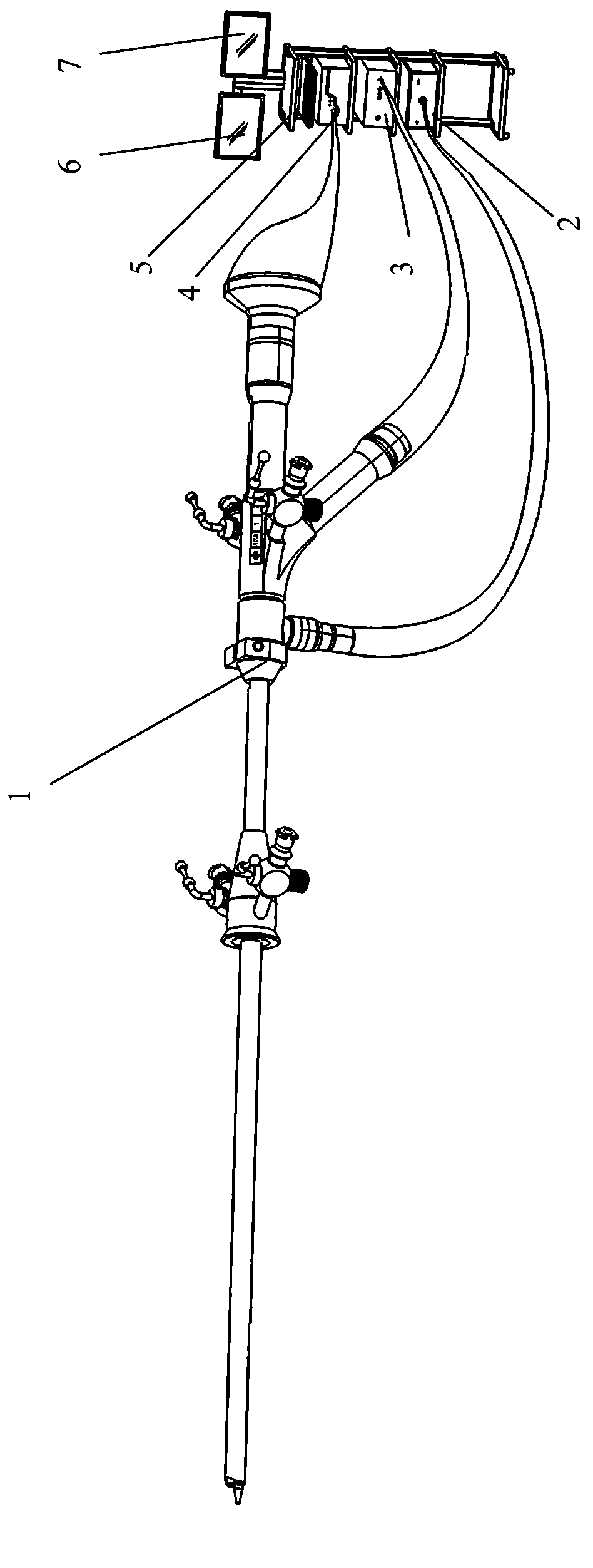
[0019] Such as figure 1 As shown, the integrated rigid ultrasonic hysteroscope system of the present invention includes an integrated rigid ultrasonic hysteroscope 1, a light source host 2, an ultrasound system host 3, a camera host 4, a keyboard 5, an endoscope monitor 6 and The micro-ultrasound monitor 7 is used to display the images of the endoscope and the micro-ultrasound of the integrated rigid ultrasonic hysteroscope 1 respectively.

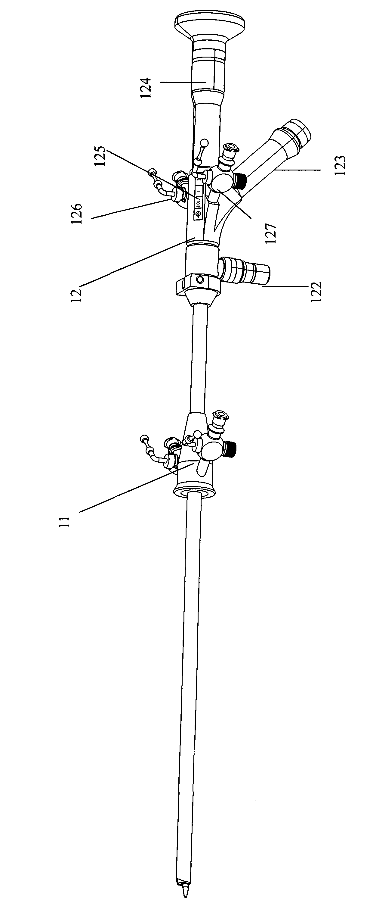
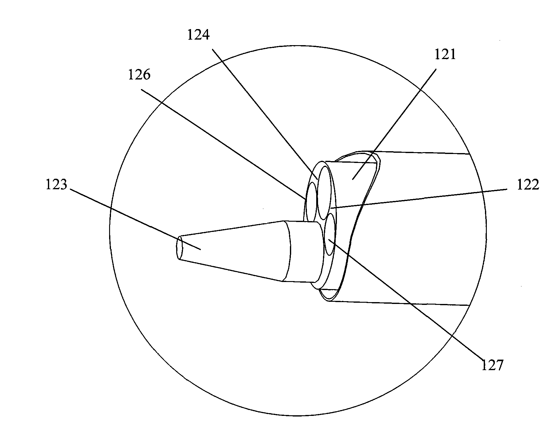
[0020] Such as figure 2 , image 3 As shown, the integrated rigid ultrasonic hysteroscope 1 described in the present invention is divided into a sheath part 11 and a main endoscope part 12 . The main body endoscope part 12 includes a rigid endoscope end 121, a light source input end 122, a miniature ultrasonic data output connector end 1123, an eyepiece input end 124, a control unit 125, two instrument channels 126 and 127, an...


PUM


Abstract

The invention belongs to the medical appliance field, in particular discloses an integral hard ultrasonic hysteroscope system. The hysteroscope system comprises a hard hysteroscope, and a light source main engine and a camera main engine which are connected with the hard hysteroscope, wherein the hard hysteroscope comprises a main endoscope part and a hysteroscope sheath part connected with the main endoscope part; the end of the hard endoscope is provided with a miniature ultrasonic probe part, an optical lens, an optical fiber and an instrument channel outlet; and the miniature ultrasonic probe part is directly integrated on the end of the hard endoscope to ensure that the hard endoscope is designed to be an integral hard ultrasonic hysteroscope. The system is characterized in that the miniature ultrasonic technique is combined with the hard hysteroscope, the miniature ultrasonic probe part is added in the structure of the hard hysteroscope, after the integral hard ultrasonic hysteroscope is placed in the uterine canal, the miniature ultrasonic probe part is started to perform linear scanning and circular scanning and obtain the true situation of the uterine canal, and the miniature ultrasonic probe part can detect the pathological changes in the uterine canal and on the uterine wall so as to help doctors to diagnose and cure. The system has good stability, the endoscope image and the ultrasonic image can be obtained more easily at the same time, the image quality in the position of lesion is increased, the diagnosis accuracy is greatly increased, and doctors are convenient to perform operation and obtain the pathological change information of the uterine canal and the uterine wall.

Description

technical field [0001] The invention belongs to the field of medical instruments, in particular to a core medical tool for gynecological surgery in modern medicine, in particular to an integrated rigid ultrasonic hysteroscope system. Background technique [0002] At present, hysteroscopy and hysteroscopy technology have been widely used clinically, and are important tools for gynecological diagnosis and treatment. Changes in the layers of the cavity and the surrounding organs have hidden dangers of insufficient diagnostic accuracy. The rigid ultrasonic hysteroscope system that has obtained the national utility model patent, its patent number is ZL200920006033.1 (announcement number: CN201299586Y), the method is to introduce micro-ultrasound technology, and the micro-ultrasound probe enters through the instrument channel of the hysteroscope Scan the uterus to obtain the conditions of the layers in the uterine cavity and between the uterine walls. The precise micro-ultrasound...

Claims


Application Information

Patent Timeline
Patent Type & Authority Applications(China)
IPC IPC(8): A61B8/12
Inventor 乔铁谢景夏黄万潮
Owner 广州市番禺区胆囊病研究所
Why Eureka
Try Eureka