Adjusting device of X-ray imaging equipment

A technology for adjusting devices and imaging equipment, which is applied in the fields of radiological diagnostic instruments, medical science, and diagnosis, and can solve problems such as the inability to change the setting of the magnification ratio, increasing the complexity of the mechanism, user operation steps, and increasing equipment costs, etc.

Active Publication Date: 2011-01-05
SHENZHEN INST OF ADVANCED TECH CHINESE ACAD OF SCI
View PDF2 Cites 29 Cited by
  • Summary
  • Abstract
  • Description
  • Claims
  • Application Information

AI Technical Summary

Problems solved by technology

[0004] In order to achieve panoramic scanning, CT scanning or head scanning, traditional imaging technology needs to use special panoramic detectors, special CT detectors or head detectors. The installation positions of each detector are fixed according to the preset magnification. The setting cannot be changed accordingly according to the real-time specific situation
The conversion of various scanning modes is completed by manually installing the corresponding detectors. The application of multiple detectors increases the cost of the equipment. Various uncertain factors such as workload and damage of each detector
The X-ray detector is fixed, it is difficult to achieve a large field of view or increase the cost
In the head scanning mode, the optical machine must be deflected at a certain angle to complete, which increases the complexity of the mechanism and the operation steps of the user

Method used


Image

  • Adjusting device of X-ray imaging equipment
  • Adjusting device of X-ray imaging equipment
  • Adjusting device of X-ray imaging equipment

Examples


Embodiment Construction

[0024] The technical solutions of the present invention will be described in detail below in conjunction with the accompanying drawings and specific embodiments.

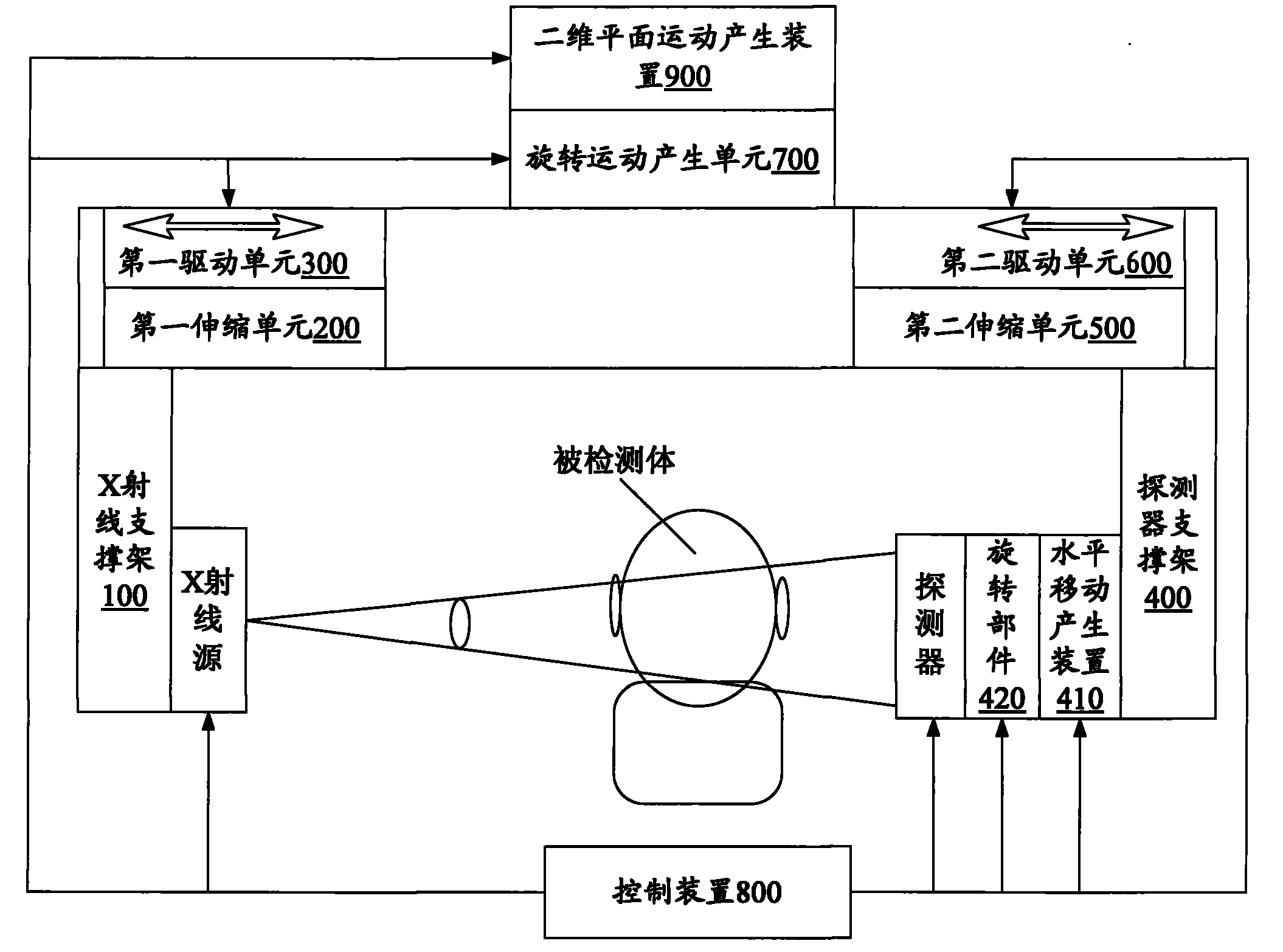
[0025] In one embodiment, such as figure 1 and figure 2 As shown, an X-ray imaging device adjustment device includes a connected X-ray support frame 100 and a first telescopic unit 200 for installing an X-ray source, and drives the X-ray support frame 100 to move along the axial direction of the first telescopic unit 200. A drive unit 300, a connected detector support frame 400 for installing the detector and a second telescopic unit 500, a second drive unit 600 that drives the detector support frame 400 to move along the axial direction of the second telescopic unit 500, and also includes a drive X The rotary motion generation unit 700 and the control device 800 for the rotation of the ray source and the detector, the rotary motion generation unit 700 is respectively socketed on the first telescopic unit 200 and ...


PUM


Abstract

The invention relates to an adjusting device of X-ray imaging equipment, comprising an X-ray supporting frame for mounting an X-ray source, a detector supporting frame for mounting a detector, a supporting beam with a sliding rail, a first driving unit and a second driving unit. The X-ray supporting frame and the detector supporting frame are arranged on the sliding rail of the supporting beam, the first driving unit drives the X-ray supporting frame to move on the sliding rail, and the second driving unit drives the detector supporting frame to move on the sliding rail. The adjusting device of the X-ray imaging equipment also comprises a control device and a rotating motion generating unit which drives the X-ray source and the detector to rotate around a rotating central shaft and is connected with the supporting beam, and the control device controls the first driving unit, the second driving unit and the rotating motion generating unit. The X-ray imaging adjusting device adjusts three distances by adopting a single detector to realize imaging scanning which integrates panorama scanning, CT (Computerized Tomography) scanning and head scanning and has simple operation and cost saving.

Description

【Technical field】 [0001] The invention relates to the field of imaging equipment, in particular to an adjustment device for X-ray imaging equipment. 【Background technique】 [0002] With the increase and aging of my country's population, the incidence of oral diseases has changed, the prevalence of dental caries has increased and the prevalence of periodontal diseases has increased, making the traditional oral health care system and resources unable to meet people's needs for oral health care and treatment. need. The development of modern medical imaging equipment provides a powerful tool for the diagnosis and treatment of oral diseases. However, ordinary domestic hospitals often use methods such as X-ray plain film and curved panoramic photography for oral examination. Although the above methods can satisfy the diagnosis and treatment of some diseases, the reliability of disease imaging diagnosis has always been affected by factors such as image overlap, deformation, and lo...

Claims


Application Information

Patent Timeline
Patent Type & Authority Applications(China)
IPC IPC(8): A61B6/00
Inventor 孙素明蒋昌辉戎军艳夏丹郑海荣
Owner SHENZHEN INST OF ADVANCED TECH CHINESE ACAD OF SCI
Why Eureka
Try Eureka