

Application of ethyl violet in DNA detection
A technology of ethyl violet and polyacrylamide gel, which is applied in the preparation of test samples, measuring devices, and the determination/inspection of microorganisms, and can solve problems such as DNA staining that have not been reported
Inactive Publication Date: 2011-04-20
WENZHOU MEDICAL UNIV
View PDF1 Cites 2 Cited by
- Summary
- Abstract
- Description
- Claims
- Application Information
AI Technical Summary
Problems solved by technology
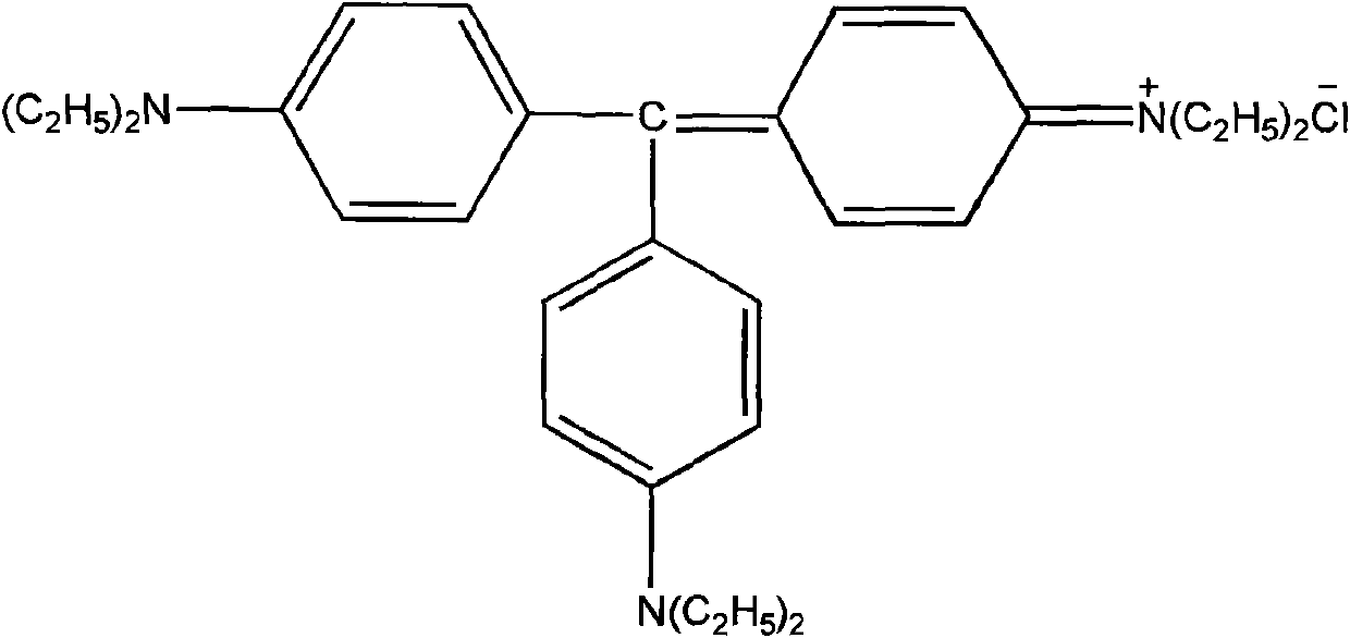
EV has been reported in the prior art for staining mammalian tissues and proteins[14-17],
Method used
View moreImage
Smart ImageViewing Examples

Examples
 PUM
 Abstract
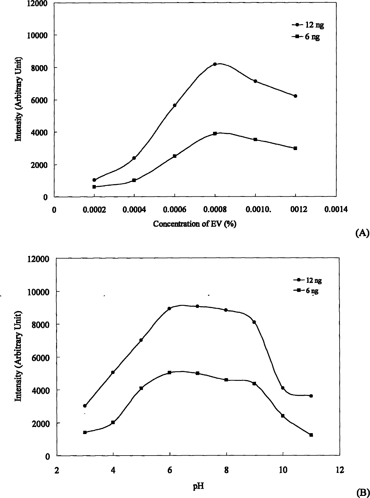
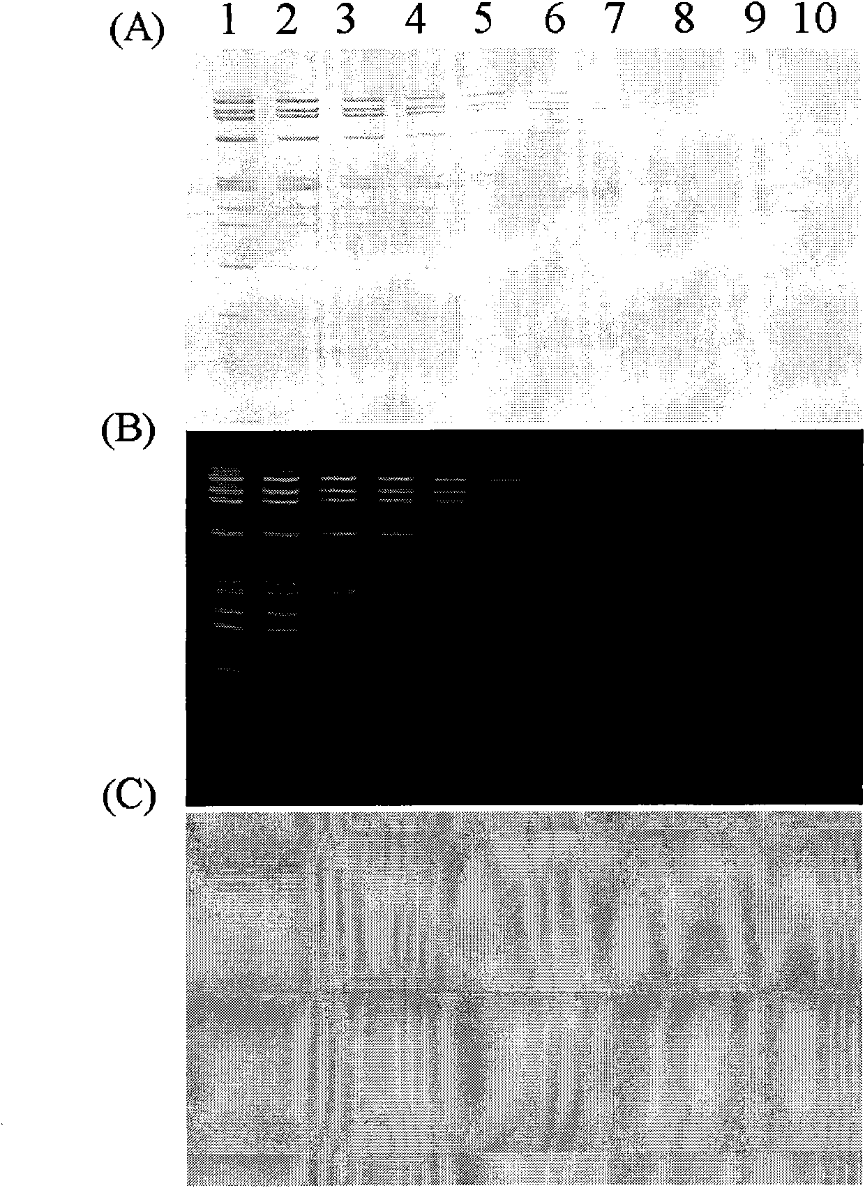
The invention relates to a method for dyeing DNA on polyacrylamide gel by using ethyl violet (EV). Dyeing solution is solution containing the ethyl violet and having the pH of 6-8. In addition, the invention also relates to a method for detecting DNA on polyacrylamide gel on the basis of the method, kits for the methods and the like.
Description
technical field [0001] The invention belongs to the technical field of biological detection. Specifically, the invention relates to a method for staining DNA with ethyl violet (EV) on a polyacrylamide gel. The background color is light. In addition, the present invention also relates to methods for detecting DNA, kits for these methods, and the like. Background of the invention [0002] Currently, electrophoresis separation of biomacromolecules such as DNA and protein on polyacrylamide gels and the coloring (visualization) of the bands have become one of the most commonly used methods in biological detection. Among them, to visualize the bands of DNA molecules on the polyacrylamide gel, isotope labeling, fluorescent dyes, visible organic dyes, silver staining and the like can be used. For reasons of sensitivity, safety, and speed, one of the most popular techniques is fluorescence detection, which uses fluorescent dyes such as ethidium bromide (EB), SYBR green, SYBR gold, ...
Claims
Login to View More Application Information
Patent Timeline
 IPC IPC(8): C12Q1/68G01N27/447G01N21/78
CPCC12Q1/6816G01N1/30C12Q2565/125C12Q2563/173C12Q2527/125
Inventor金利泰丛维涛李校堃梁广冯治国林绍强朱忠欣张美玲
OwnerWENZHOU MEDICAL UNIV