Method for automatically generating liver 3D (three-dimensional) image and accurately positioning liver vascular domination region

An accurate positioning and automatic generation technology, applied in the physical field, can solve the problems of inaccurate liver range and limited means

An accurate positioning and automatic generation technology, applied in the physical field, can solve the problems of inaccurate liver range and limited means

CN102048550BInactive Publication Date: 2013-07-17RENJI HOSPITAL AFFILIATED TO SHANGHAI JIAO TONG UNIV SCHOOL OF MEDICINE

Image

  • Method for automatically generating liver 3D (three-dimensional) image and accurately positioning liver vascular domination region
  • Method for automatically generating liver 3D (three-dimensional) image and accurately positioning liver vascular domination region
  • Method for automatically generating liver 3D (three-dimensional) image and accurately positioning liver vascular domination region

Examples


Embodiment Construction

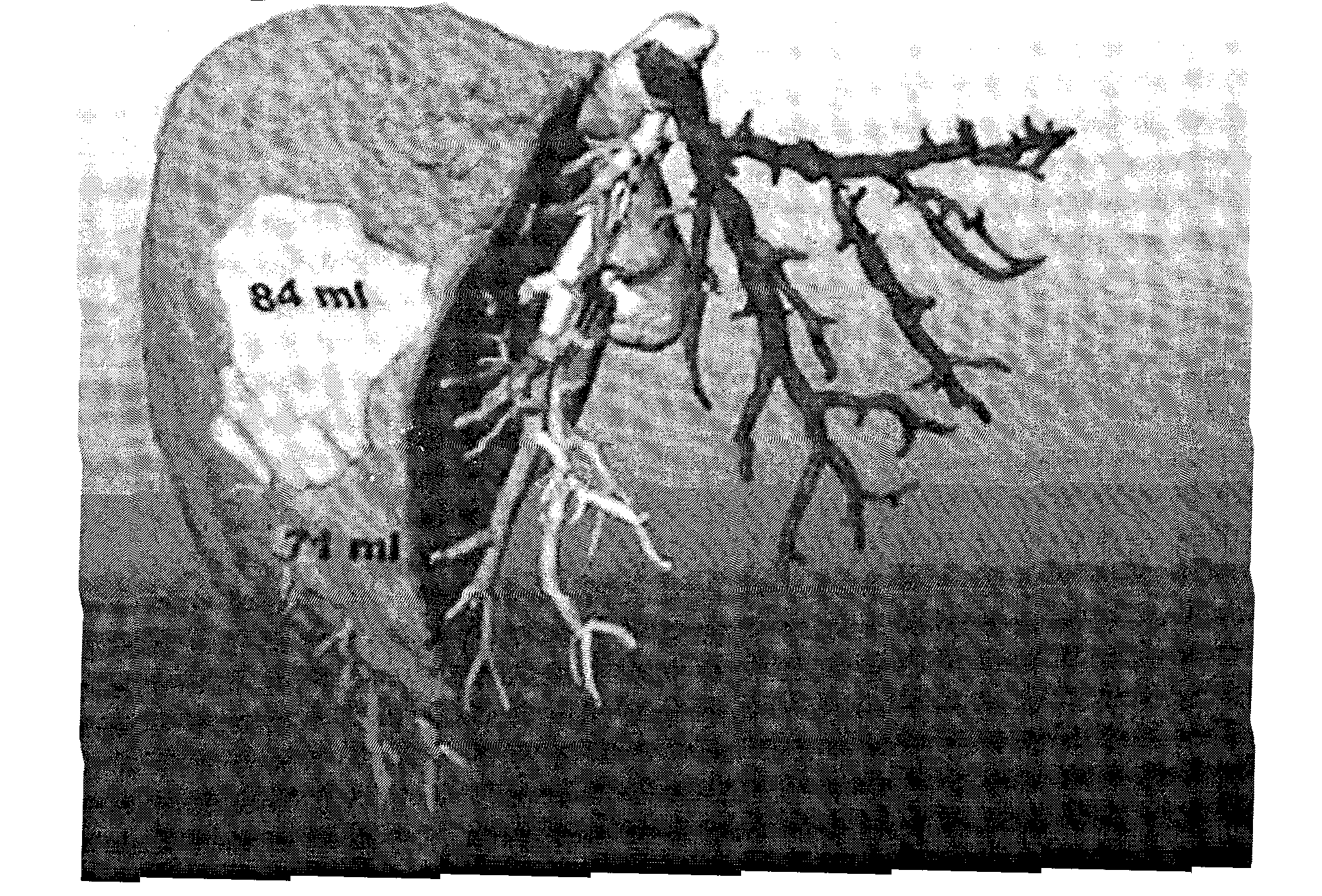
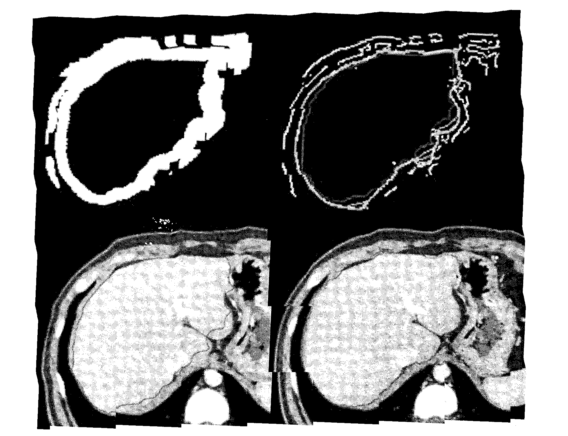
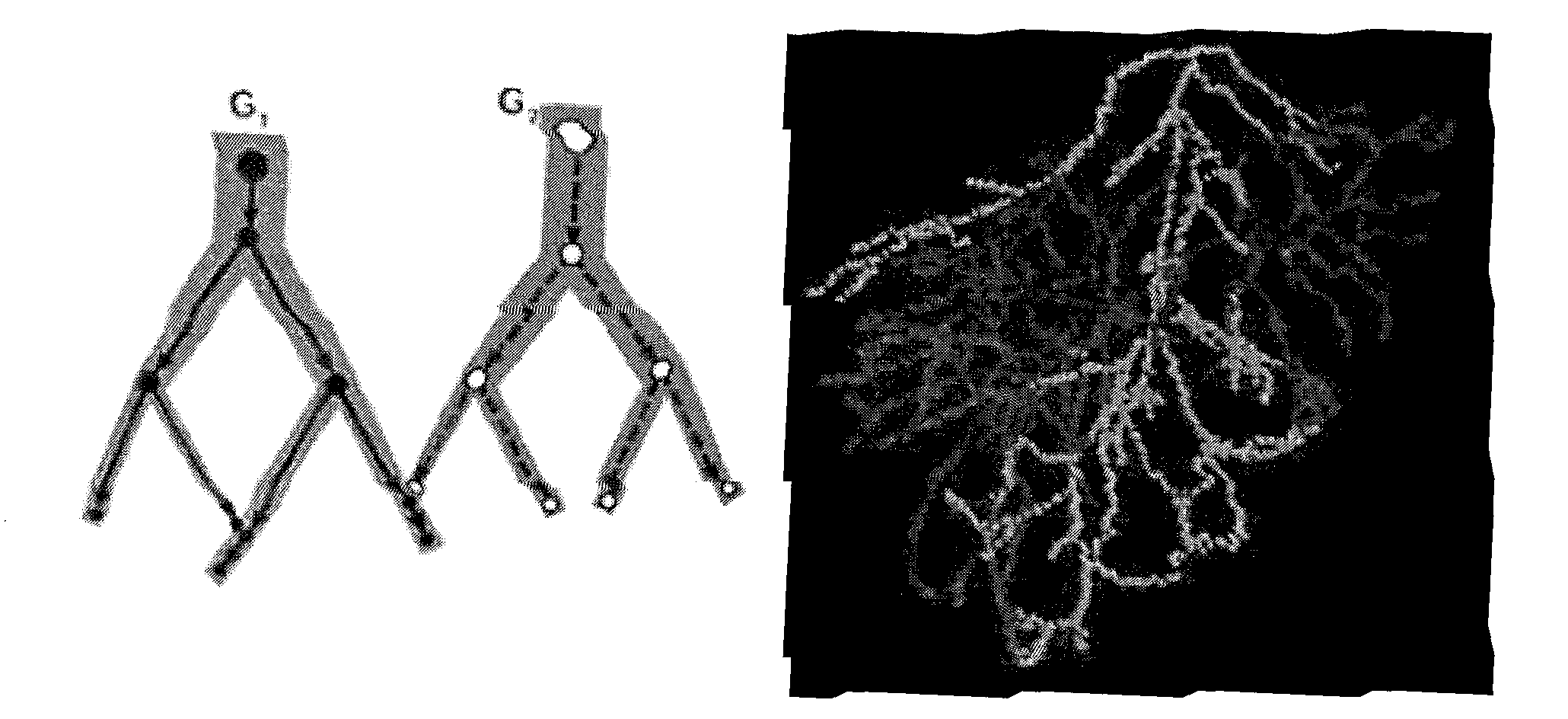
[0025] Such as Figure 5 As shown, the present invention provides a method for automatically generating a liver 3D image and accurately locating the hepatic vascular dominance area. The method includes a process of obtaining a three-dimensional CT enhanced image of the liver, including a process of locating and The process of segmenting the liver includes a process of extracting liver blood vessels in the image and analyzing its structure, including a process of analyzing the areas dominated by blood vessels.

[0026] specific,

[0027] a) In the process of obtaining a three-dimensional CT enhanced image of the liver, a tomographic scanner is used to obtain a tomographic image of the liver, and a multi-row spiral CT contrast agent is used to perform enhanced three-phase dynamic scanning of the human body to obtain the original data of the liver, and then inject the contrast agent into the human body Afterwards, conventional upper abdominal CT enhanced three-phase dynamic scan...


PUM


Abstract

The invention provides a method for automatically generating a liver 3D (three-dimensional) image and accurately positioning a liver vascular domination region. The method comprises the following processes: acquiring a liver three-dimensional CT (computed tomography)-enhanced image; positioning and segmenting the liver; extracting liver blood vessels and analyzing the structure of the liver; and analyzing the vascular domination region. In the method, through the topology partitioning, the volume size of the drainage region of each venous blood vessel can be obtained so as to facilitate a surgeon to perform simulation and prediction before a liver surgery, and simultaneously facilitate research and understanding of positions of blood vessels in the liver region for scientific researchers.

Description

Technical field: [0001] The invention relates to the field of physics, in particular to graphic measurement and processing technology, in particular to a method for automatically generating a 3D image of the liver and accurately locating the dominating area of ​​liver vessels. Background technique: [0002] With the advent of the era of precise liver resection and living donor liver transplantation, precise liver segment surgery is the development trend of hepatobiliary surgery, which requires accurate preoperative estimation and measurement of the resected liver segment. The accuracy of CT for liver volume measurement has been recognized and even considered the gold standard. However, surgical operations often need to know the size of the liver volume in the blood supply of a certain portal vein or the drainage range of the hepatic vein, so as to judge how much blood flow in the liver will be affected after processing these blood vessels. However, since there are no specia...

Claims


Application Information

Patent Timeline
17 Jul 2013
Publication
CN102048550B
IPC
A61B6/03; G06F19/00; G06T15/00
Inventors
许建荣; 钱黎俊