Combined rigid ureteroscope

A ureteroscope and combined technology, applied in the field of ureteroscope, can solve the problems of injury risk, increase patient pain, prolong operation time, etc., and achieve the effect of avoiding difficult operation and easy damage, improving operation safety and clinical treatment safety

Active Publication Date: 2011-05-25
周均洪
View PDF4 Cites 15 Cited by
  • Summary
  • Abstract
  • Description
  • Claims
  • Application Information

AI Technical Summary

Problems solved by technology

When the existing ureteroscope structure is inspected and lithotripsy, the stones need to be taken out of the body through the lumen of the ureteroscope after being crushed by the lithotripsy instrument. Because the lumen is very small, slightly larger gravels cannot pass through. In this way, the ureteroscope needs to repeatedly enter and exit the urethra, bladder, ureter, and even the kidney to remove stones, which will prolong the operation time and increase the operation risk, and the repeated entry and exit will also increase the pain of the patient; On the one hand, in the application of flexible ureteroscope to the diagnosis and treatment of renal pelvis and calyx and upper ureteral diseases, it is first necessary to use rigid ureteroscope to dilate the ureteral orifice and perform microscopic examination, and indwell the guide wire, then withdraw the rigid ureteroscope and follow the guide wire Put the flexible tube into the ureter, pull out the guide wire, and place a flexible ureteroscope along the flexible ureter to diagnose and treat diseases of the upper ureter, renal pelvis, and calices. Since the flexible tube cannot be placed under direct vision during the operation, It will increase the risk of ureteral injury, and the operation is complicated. The flexible ureteroscope is not only expensive, difficult to operate and easy to wear and tear, but its frequent use will result in long operation time and high operation cost, which is not easy to popularize and carry out. Therefore, the existing The structural design of the ureteroscope cannot meet the requirements of safe, effective and cheap modern clinical treatment

Method used


Image

  • Combined rigid ureteroscope
  • Combined rigid ureteroscope
  • Combined rigid ureteroscope

Examples


Embodiment Construction

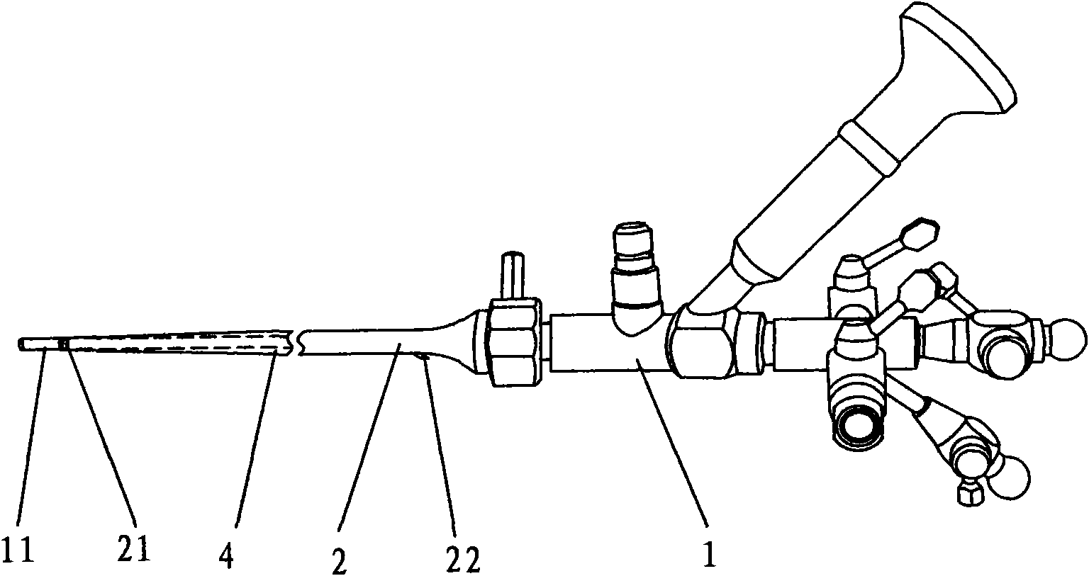
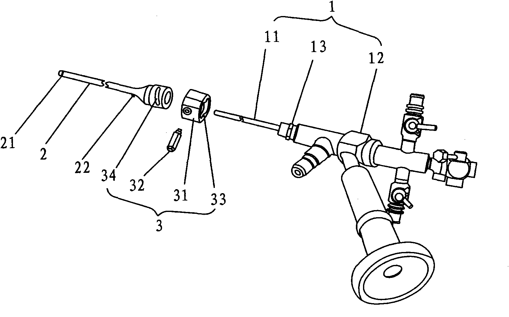
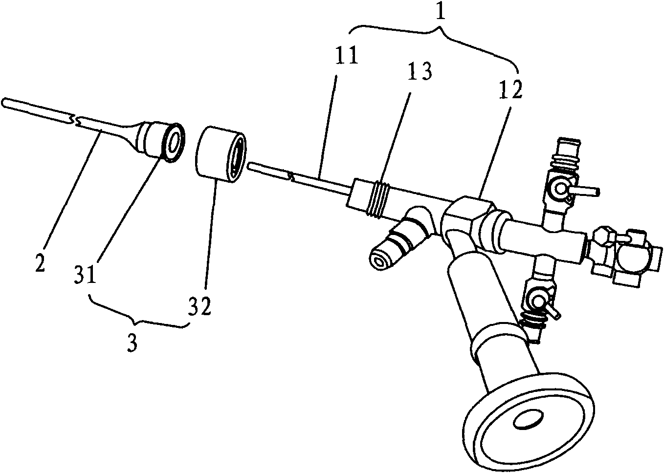
[0017] refer to figure 1 , figure 2 , a combined rigid ureteroscope, comprising a rigid ureteroscope body 1 and a tubular scope sheath 2 set outside the scope tubular portion 11, the starting end of the scope sheath 2 is closely attached to the scope tubular portion 11 Close, the rear end of the mirror sheath 2 is provided with a locking mechanism 3, a locking part 13 is provided between the tubular part 11 of the mirror body and the operating part 12, and the locking mechanism 3 is matched with the locking part 13 Lock or loosen the rigid ureteroscope body 1 and the tubular scope sheath 2.

[0018] In this embodiment, the locking mechanism 3 at the rear end of the mirror sheath 2 includes a sleeve 31 with an end cap 33 and a pin 32 fixed on the side wall of the sleeve 31, and the outer wall of the rear end of the tubular mirror sheath 2 An inclined chute 34 is provided, and the end of the dial pin 32 extends into the chute 34; the locking part 13 of the mirror body 1 is an...


PUM


Abstract

The invention discloses a combined rigid ureteroscope which comprises a rigid ureteroscope body and a tubular sheath which is sheathed outside a tubular part of the ureteroscope body, wherein a starting end of the sheath is tightly attached to the tubular part of the ureteroscope body; the rear end of the sheath is equipped with a locking mechanism; a locking part is arranged between the tubular part and an operation part of the ureteroscope body; and the locking mechanism is matched with the locking part so as to lock or loosen the rigid ureteroscope body and the tubular sheath. By adopting the combined rigid ureteroscope,, the locking mechanism between the ureteroscope body and the sheath can be loosened during operations for examination, diagnosis and treatment of ureter and kidney, the rigid ureteroscope body and the sheath can be separated, and the sheath is left in the ureter and utilized as a channel through which broken stones can be repeatedly taken out for multiple times or the ureteroscope body separately gets in and out of the ureter as well as an operation channel of a flexible ureteroscope, thereby saving the operation procedures, shortening the operation time and improving the operation safety; and moreover, in some operation procedures, the rigid ureteroscope can be used instead to implement projects that must be implemented by the flexible ureteroscope before,thereby avoiding the problems of difficult operation and easy damage of the flexible ureteroscope, and actually realizing safe, effective and low-cost clinical treatment.

Description

technical field [0001] The invention relates to a medical device, in particular to a ureteroscope. Background technique [0002] Ureteroscope is an endoscope used for the inspection, diagnosis and treatment of ureter and kidney. It includes soft ureteroscope and rigid ureteroscope, which can be adapted to the diagnosis and treatment of different parts of the upper urinary tract according to their characteristics. . When the existing ureteroscope structure is inspected and lithotripsy, the stones need to be taken out of the body through the lumen of the ureteroscope after being crushed by the lithotripsy instrument. Because the lumen is very small, slightly larger gravels cannot pass through. In this way, the ureteroscope needs to repeatedly enter and exit the urethra, bladder, ureter, and even the kidney to remove stones, which will prolong the operation time and increase the operation risk, and the repeated entry and exit will also increase the pain of the patient; On the...

Claims


Application Information

Patent Timeline
Patent Type & Authority Applications(China)
IPC IPC(8): A61B1/307A61B1/015
CPCA61B17/3421A61B1/00105A61B1/307A61B2017/345A61B2017/00477A61B1/015A61B17/22A61B1/00135A61B1/00078
Inventor 周均洪
Owner 周均洪
Why Eureka
Try Eureka