Triple differential confocal fundus retina scanning and imaging device and method on basis of adaptive optics

An adaptive optics, differential confocal technology, applied in the fields of ophthalmoscope, application, medical science, etc., can solve the problem of failing to improve the lateral resolution capability of confocal microscope, high operating environment requirements, and difficulty in ensuring pinhole displacement. stability issues

Active Publication Date: 2012-11-07
INST OF OPTICS & ELECTRONICS - CHINESE ACAD OF SCI
View PDF2 Cites 0 Cited by
  • Summary
  • Abstract
  • Description
  • Claims
  • Application Information

AI Technical Summary

Problems solved by technology

However, due to its non-common optical path structure, it is difficult to ensure the stability of the pinhole displacement, and the requirements for the use environment are relatively high.
And this method failed to improve the lateral resolving power of the confocal microscope
At the same time, the low light energy utilization efficiency of this method in low-light detection limits its application in the field of in vivo retinal imaging.

Method used


Image

  • Triple differential confocal fundus retina scanning and imaging device and method on basis of adaptive optics
  • Triple differential confocal fundus retina scanning and imaging device and method on basis of adaptive optics
  • Triple differential confocal fundus retina scanning and imaging device and method on basis of adaptive optics

Examples


Embodiment Construction

[0032] specific implementation plan

[0033] The embodiment of the present invention combines adaptive optics confocal scanning ophthalmoscope technology and three-differential confocal microscope technology to obtain high-resolution live retinal images, as follows:

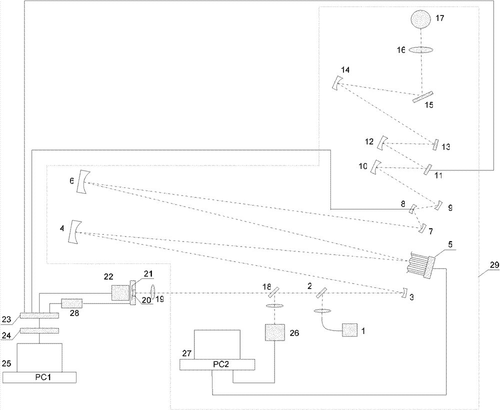
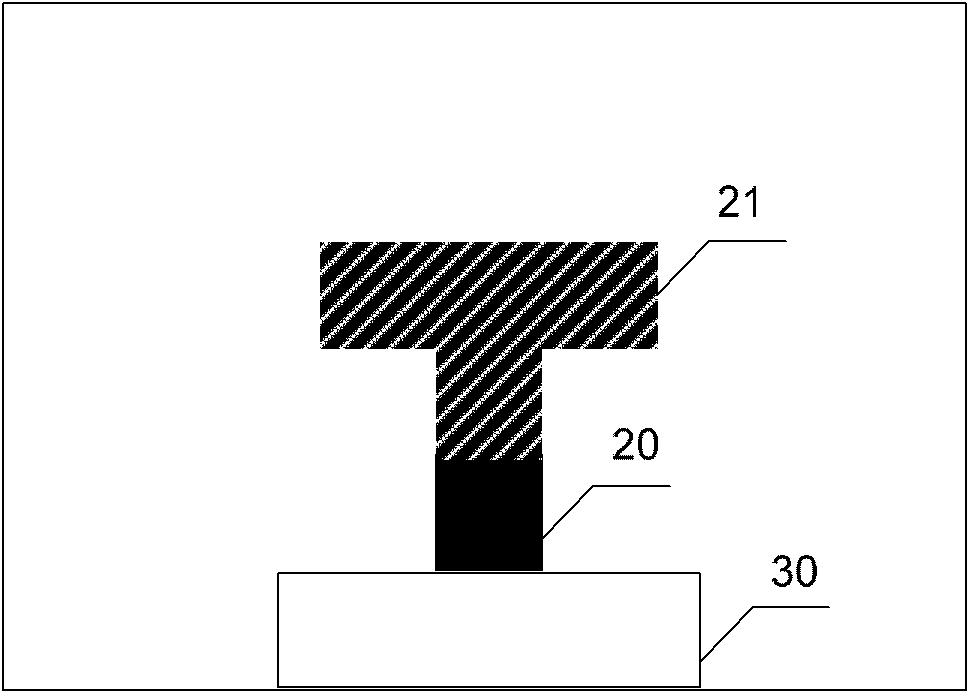
[0034] Such as figure 1 As shown, the imaging device of the present invention includes: an optical system and an adaptive optics control system 29 based on an adaptive optics laser confocal ophthalmoscope (AOSLO), a pinhole axial micro-displacement driving device 28, a pinhole axial micro-displacement Device 20, light detection place pinhole 21, light detection device 22, signal synchronization device 23, data acquisition device 24 and data processing device 25; The optical system and automatic The adaptive optics control system 29 includes a light source 1, a first spherical reflector 3, a second spherical reflector 4, a deformable mirror 5, a third spherical reflector 6, a fourth spherical reflector 7, a fift...


PUM


Abstract

The invention relates to a triple differential confocal fundus retina scanning and imaging device and method on the basis of adaptive optics. On the basis of the adaptive optics confocal scanning laser ophthalmoscope (AOSLO) of the adaptive optics, a triple differential detecting technique is utilized to further increase the axial resolution ratio of AOSLO and realize high resolution imaging of the fundus retina. In a system light path, a pinhole axial micro-displacement device is utilized to change the axial position of a pinhole in an optical detection part and realize the triple differential probing of signals. The invention relates to a high resolution fundus retina imaging method and device which have the advantages of simplicity and efficient utilization of optical energy.

Description

technical field [0001] The invention relates to a device and method capable of high-resolution imaging of the retina, in particular to a high-resolution imaging method and imaging device based on adaptive optics-based three-differential confocal scanning retinal fundus. Background technique [0002] The laser confocal scanning microscope places a point light source, a point object, and a point detector at positions conjugate to each other, and uses scanning to realize imaging. Its basic principle is as image 3 As shown, the light emitted by the point light source 31 passes through the objective lens 32 and the half-mirror 33 and is focused into a point on the observation sample surface 34 and reflected, and the reflected light returns along the original path, and then passes through the half-mirror 33 and the collecting lens 35 The signal light from the observation sample surface 34 is guided into the pinhole 36 and is detected at a photodetector 37 . Only the signal light...

Claims


Application Information

Patent Timeline
Patent Type & Authority Patents(China)
IPC IPC(8): A61B3/12A61B3/14
CPCA61B3/10A61B3/1025
Inventor 张雨东李昊卢婧史国华
Owner INST OF OPTICS & ELECTRONICS - CHINESE ACAD OF SCI
Why Eureka
Try Eureka