Computer-aided method for distinguishing ultrasound endoscope image of pancreatic cancer

A computer-aided, ultrasonic image technology, applied in the field of medical instruments, can solve problems such as large impact, different diagnostic accuracy, and difficult images, and achieve the effect of improving accuracy

Inactive Publication Date: 2011-07-13
SECOND MILITARY MEDICAL UNIV OF THE PEOPLES LIBERATION ARMY
View PDF2 Cites 14 Cited by
  • Summary
  • Abstract
  • Description
  • Claims
  • Application Information

AI Technical Summary

Problems solved by technology

However, the diagnosis based on endoscopic ultrasound images is greatly affected by the doctor's experience and subjective factors, and the diagnostic accuracy of different doctors will be different; some subt

Method used


Image

  • Computer-aided method for distinguishing ultrasound endoscope image of pancreatic cancer
  • Computer-aided method for distinguishing ultrasound endoscope image of pancreatic cancer

Examples


Embodiment Construction

[0026] Below in conjunction with specific embodiment, further illustrate the present invention. It should be understood that these examples are only used to illustrate the present invention and are not intended to limit the scope of the present invention. In addition, it should be understood that after reading the teachings of the present invention, those skilled in the art can make various changes or modifications to the present invention, and these equivalent forms also fall within the scope defined by the appended claims of the present application.

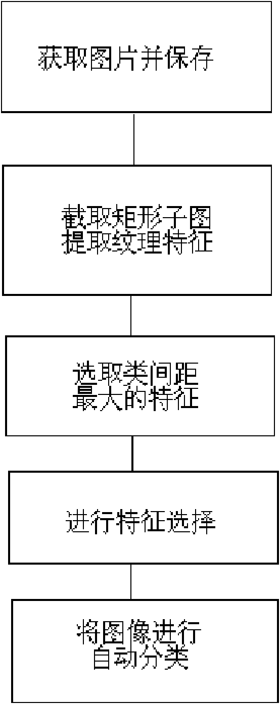
[0027] The present invention comprises the following steps:

[0028] (1) Place an endoscope on the top of the miniature high-frequency ultrasonic probe to obtain an endoscopic ultrasonic image, and save it in the form of Windows bitmap format BMP;

[0029] (2) According to the ROIs, intercept the rectangular submap, extract the texture features used in the classification of pancreas EUS images in each ROIs, and screen them by ...


PUM


Abstract

The invention relates to a computer-aided method for distinguishing the ultrasound endoscope image of pancreatic cancer, providing a method for extracting and classifying the textural features of the ultrasound endoscope image of pancreatic cancer. The computer-aided method can be used for the computer-aided analysis of the ultrasound endoscope image of pancreatic cancer. 9 general classes which totally comprise 69 textural features are extracted from the ultrasound endoscope image of pancreatic cancer by a digital image processing algorithm. Class spacing is adopted to serve as a separability criterion to preliminarily screen the features; then, a chronological progress search algorithm is used for further screening the features; and the features are classified by a support vector machine. The method is realized by extracting the textural features of the ultrasound endoscope image via the classifier, various objective quantized diagnostic indexes and a method for correctly describing and explaining the ultrasound endoscope image are built, and the accuracy on the ultrasound endoscope early diagnosis of the pancreatic cancer is improved.

Description

technical field [0001] The invention belongs to the technical field of medical instruments, in particular to a computer-aided method for discriminating ultrasonic endoscopic images of pancreatic cancer. Background technique [0002] Pancreatic cancer is one of the top ten malignant tumors announced by the World Health Organization, and it is a major disease that seriously endangers human health. In recent years, the incidence of pancreatic cancer has been increasing year by year. Pancreatic cancer develops rapidly and is dangerous, and its clinical features are short course of disease, rapid progression, and high mortality. [0003] The pancreas is a retroperitoneal organ with a hidden location, complex adjacency, and many important organs and blood vessels around it. Therefore, the early detection and early diagnosis of pancreatic cancer have long been a difficult problem in the medical field. [0004] Endoscopic uhrasonography (EUS) has been widely used all over the wor...

Claims


Application Information

Patent Timeline
IPC IPC(8): G06K9/62G06K9/00
Inventor 张敏敏金震东李兆申蔡哲元吴仪俊余建国
Owner SECOND MILITARY MEDICAL UNIV OF THE PEOPLES LIBERATION ARMY
Why Eureka
Try Eureka