Method for measuring femoral head-neck spatial angles

A space angle and measurement method technology, applied in computer tomography scanners, echo tomography, etc., can solve the problems of inability to observe the state of femur space angle intuitively, and achieve the effect of convenient measurement, fast and convenient collection, and clear display

Inactive Publication Date: 2011-08-17
THE FIRST AFFILIATED HOSPITAL OF THIRD MILITARY MEDICAL UNIVERSITY OF PLA
View PDF3 Cites 13 Cited by
  • Summary
  • Abstract
  • Description
  • Claims
  • Application Information

AI Technical Summary

Problems solved by technology

[0004] The method for measuring the spatial angle of the femur in the above scheme has the following disadvantages: it is all measured on two-dimensional images, and it is difficult to intuitively measure various spatial angles of the femur, and the final generated image is also a two-dimensional image, which cannot be intuitively measured. The observation of various spatial angle states of the femur

Method used


Image

  • Method for measuring femoral head-neck spatial angles
  • Method for measuring femoral head-neck spatial angles
  • Method for measuring femoral head-neck spatial angles

Examples


Embodiment Construction

[0019] The known structures and methods in this solution will not be described here. Follow the steps below:

[0020] (1) Place the complete human skeleton specimen on the scanning table in a normal state, with the midsagittal line of the human body parallel to the midline of the examination table.

[0021] (2) CT scanning: use multi-slice spiral CT scanning to continuously collect data on the human body to obtain thin-slice sequences. The scanning parameters are 120KV and 150MAS, the slice thickness is 1 mm, and the interval is 0.6 mm. The scanning range is from the upper edge of the acetabulum to the lower to the lesser trochanter of the femur.

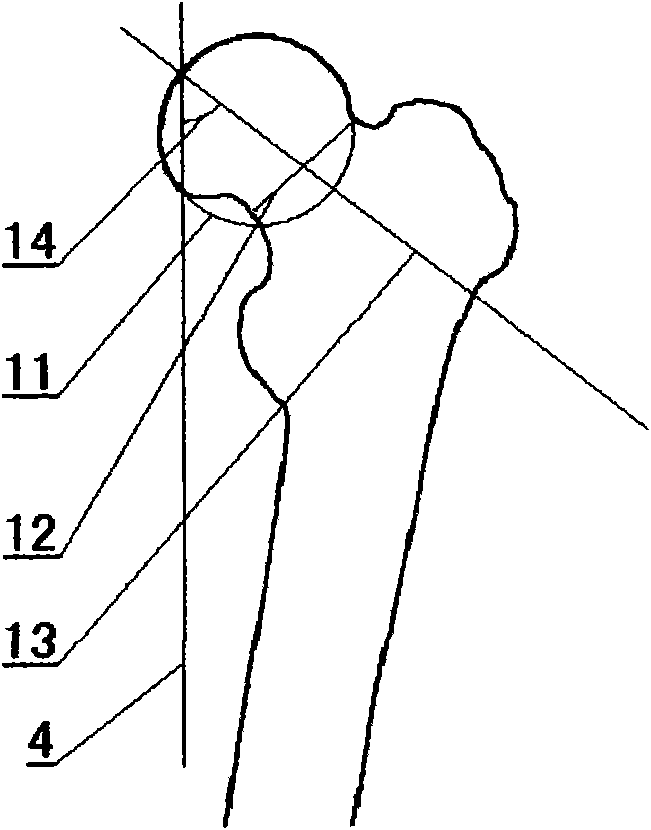
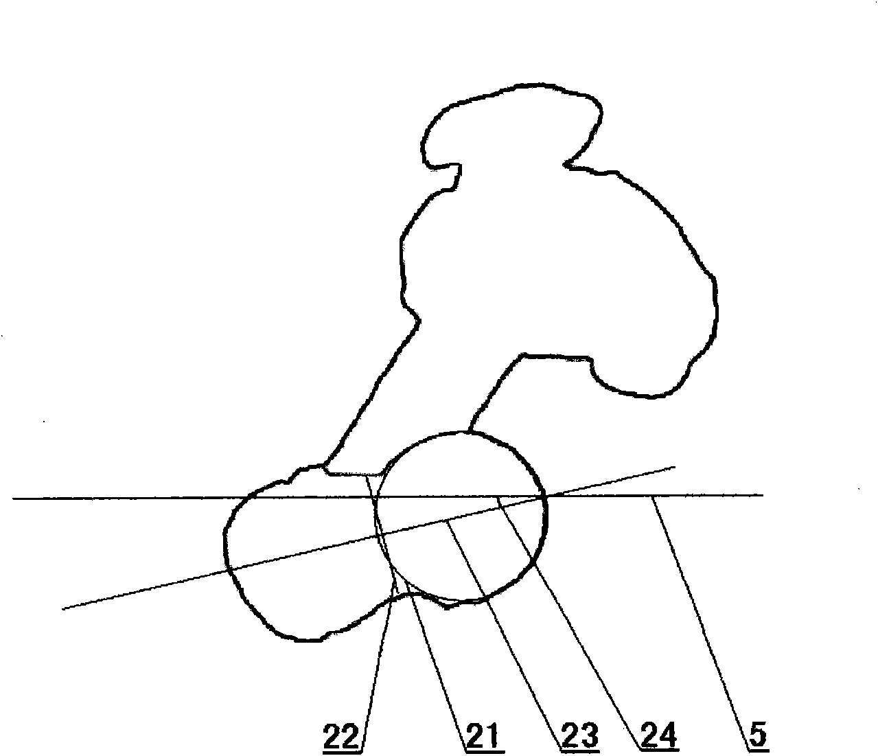
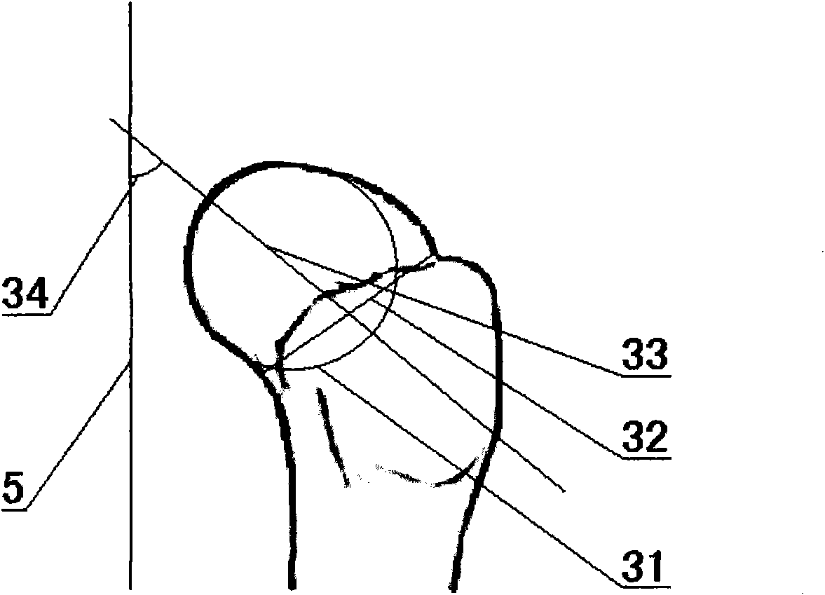
[0022] (3) Three-dimensional reconstruction: using a three-dimensional post-processing program to reconstruct a three-dimensional image of the hip joint from the thin-slice sequence, removing irrelevant structures such as the acetabulum, and obtaining a three-dimensional image of the femur.

[0023] (4) The three-dimensional image...


PUM


Abstract

The invention relates to a measuring method for CT (Computed Tomography) scanning and postprocessing and aims to provide a three-dimensional method for measuring femoral head-neck spatial angles. The method comprises the following steps: enabling a measured person to lie on the back on an examination bed, and enabling the middle sagittal line of the human body to coincide with the middle line of the examination bed; carrying out continuous data acquisition on the human body by adopting multilayer spiral CT scanning to obtain a thin-layer sequence, wherein the scanning range is from the upper edge of acetabulum to the lesser trochanter of femur; reconstructing a hip joint three-dimensional image based on the thin-layer sequence by using a three-dimensional postprocessing program, and removing unrelated structures such as the acetabulum and the like to obtain a femur three-dimensional image; and measuring the femoral head-neck extraversion angle, postversion angle and subversion angle. The technical scheme of the invention has the characteristic that the surface anatomical structure of the three-dimensionally restructured femur is displayed clearly, the position can be easily regulated, and the femoral head-neck spatial angles can be precisely measured.

Description

technical field [0001] The invention relates to a measurement method of CT scanning and post-processing, in particular to a method for measuring the spatial angle of the femoral head and neck by using a three-dimensional post-processing image. Background technique [0002] The measurement of the space angle of the femoral head and neck has a guiding role in the reduction of femoral head and neck fractures and the manufacture and installation of artificial hip joints, and has very important clinical significance. [0003] Nowadays, the measurement of human femoral space angle is mainly through CT method and bare bone projection measurement method (hereinafter referred to as "bare bone method"). The typical CT method is to place the specimen flat on the scanning bed, and measure the femoral head and neck perpendicular to the long axis of the femur. and femoral condyle level for two-dimensional scanning. The typical bare-bone method is to place the femoral specimen on a horizo...

Claims


Application Information

Patent Timeline
Patent Type & Authority Applications(China)
IPC IPC(8): A61B6/03
Inventor 戴明德王健陈伟蔡萍周代全黎川陆明黄学全陈丽陈莉程琳陈霖
Owner THE FIRST AFFILIATED HOSPITAL OF THIRD MILITARY MEDICAL UNIVERSITY OF PLA
Why Eureka
Try Eureka