Cardiac- and/or respiratory-gated image acquisition system and method for virtual anatomy enriched real-time 2D imaging in interventional radiofrequency ablation or pacemaker placement procedures

A technology of image acquisition and anatomical structure, which is applied in the direction of radiological diagnosis equipment, electrocardiography, application, etc., can solve the problems of inaccurate registration accuracy and failure to consider the patient's breathing and heart movement

Active Publication Date: 2011-09-21
KONINK PHILIPS ELECTRONICS NV
View PDF6 Cites 25 Cited by
  • Summary
  • Abstract
  • Description
  • Claims
  • Application Information

AI Technical Summary

Problems solved by technology

On the other hand, such registration does not take into account the breathing and cardiac motion of the patient, which may lead to the fact that the obtained registration accuracy is not precise enough for some applications

Method used


Image

  • Cardiac- and/or respiratory-gated image acquisition system and method for virtual anatomy enriched real-time 2D imaging in interventional radiofrequency ablation or pacemaker placement procedures
  • Cardiac- and/or respiratory-gated image acquisition system and method for virtual anatomy enriched real-time 2D imaging in interventional radiofrequency ablation or pacemaker placement procedures
  • Cardiac- and/or respiratory-gated image acquisition system and method for virtual anatomy enriched real-time 2D imaging in interventional radiofrequency ablation or pacemaker placement procedures

Examples


Embodiment Construction

[0030] In the following, the proposed image acquisition device and method according to the present invention will be explained in more detail with respect to particular refinements and with reference to the accompanying drawings.

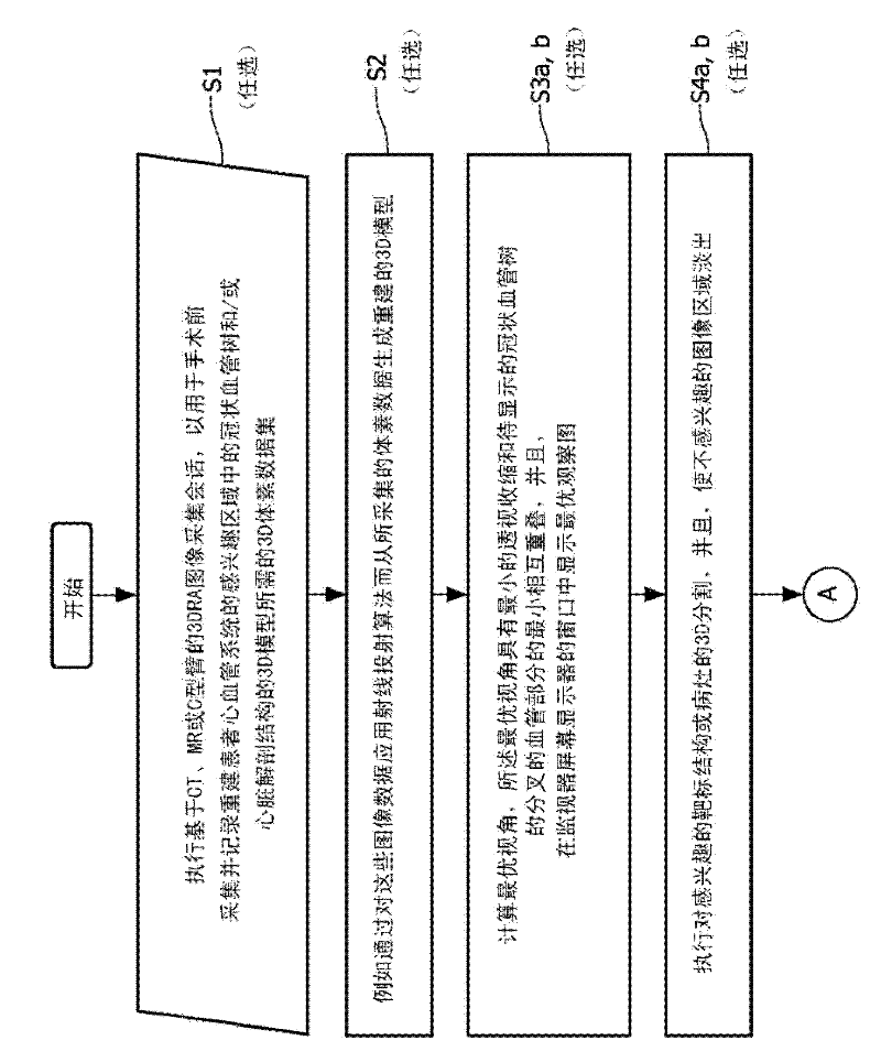
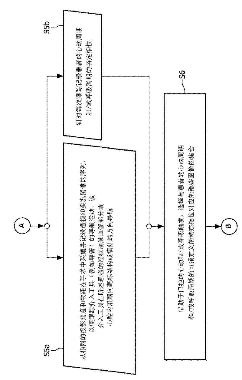
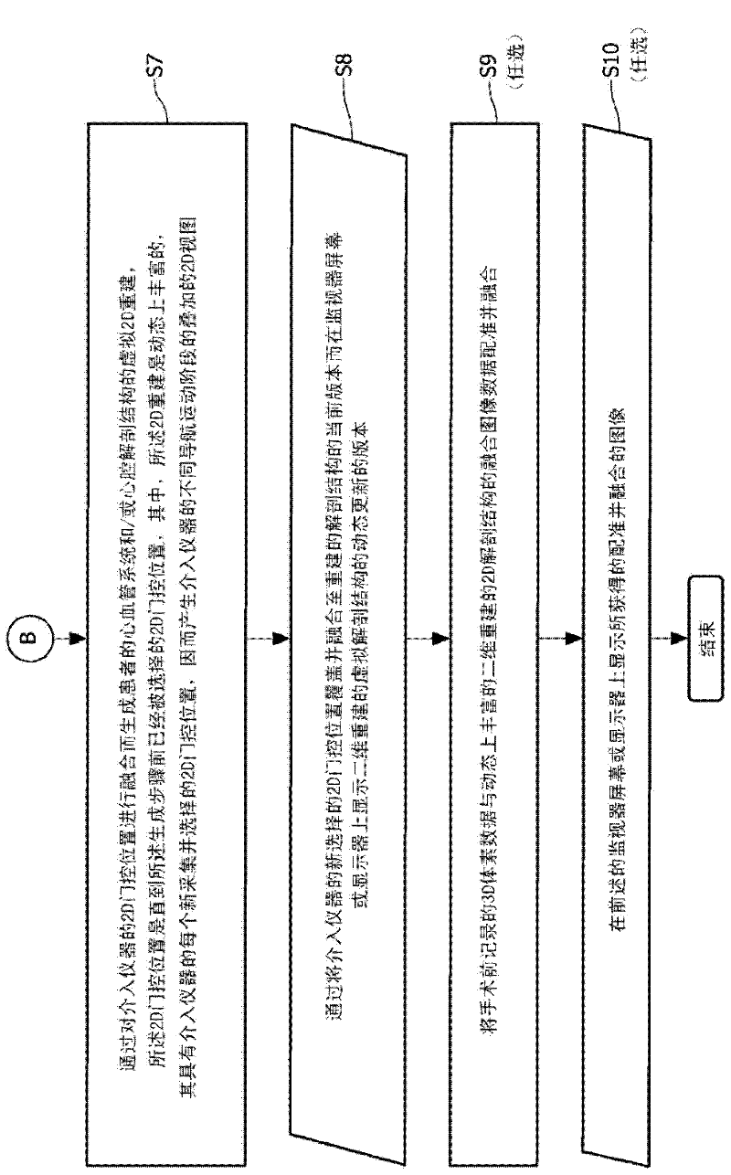
[0031] The flowchart depicted in Fig. 1 illustrates the proposed image acquisition method according to the above-described first exemplary embodiment of the present invention. As already mentioned above, the proposed method starts with the optional step of performing an image acquisition session based on CT, MR or C-arm 3DRA or any other modality type (ultrasound, imaging, etc.) to A 3D voxel data set required to reconstruct a 3D model of the coronary vascular tree and / or cardiac anatomy in a region of interest of the patient's cardiovascular system and record (S1) for the preoperative acquisition and recording (S1) of the preoperative image data acquisition step Thereafter, a three-dimensional reconstructed model or a 3D map of the patient's cardio...


PUM


Abstract

The present invention refers to the field of cardiac electrophysiology (EP) and, more specifically, to image-guided radio frequency ablation and pacemaker placement procedures. For those procedures, it is proposed to display the overlaid 2D navigation motions of an interventional tool intraoperatively obtained from the same projection angle for tracking navigation motions of an interventional tool during an image-guided intervention procedure while being navigated through a patient's bifurcated coronary vessel or cardiac chambers anatomy in order to guide e.g. a cardiovascular catheter to a target structure or lesion in a cardiac vessel segment of the patient's coronary venous tree or to a region of interest within the myocard. In such a way, a dynamically enriched 2D reconstruction of the patient's anatomy is obtained while moving the interventional instrument. By applying a cardiac and / or respiratory gating technique, it can be provided that the 2D live images are acquired during the same phases of the patient's cardiac and / or respiratory cycles. Compared to prior-art solutions which are based on a registration and fusion of image data independently acquired by two distinct imaging modalities, the accuracy of the two-dimensionally reconstructed anatomy is significantly enhanced.

Description

technical field [0001] The present invention relates to the field of cardiac electrophysiology (EP), and, more particularly, to image-guided radiofrequency ablation and pacemaker placement procedures. For those procedures, it is proposed that the interventional tool navigate through the patient's bifurcated coronary vessels or cardiac cavity anatomy in order to guide, for example, a cardiovascular catheter to a target structure or lesion in the cardiovascular portion of the patient's coronary venous tree or to the myocardium When within the region of interest, display the overlaid 2D navigation motion of the interventional tool from the same projection angle as acquired during surgery. In this way, a dynamically rich 2D reconstruction of the patient's anatomy is obtained while moving the interventional instrument. By applying cardiac and / or respiratory gating techniques, acquisition of 2D live images during the same phase of the patient's cardiac cycle and / or respiratory cycl...

Claims


Application Information

Patent Timeline
Patent Type & Authority Applications(China)
IPC IPC(8): A61B6/03A61B5/0456A61B5/352
CPCA61B8/5276A61B19/5225A61B2019/5289A61B6/504A61B6/5235A61B6/5247A61B8/0891A61B6/12A61B6/527A61B6/5288A61B6/032A61B5/0456A61B5/1135A61B5/7289A61B6/503A61B2090/364A61B90/37A61B5/352
Inventor N·P·B·戈金C·A·M·皮卡尔N·F·维兰
Owner KONINK PHILIPS ELECTRONICS NV
Why Eureka
Try Eureka