Method and apparatus for measuring pneumothorax

A technology for pneumothorax and thorax, which is applied in the medical field and can solve the problems of not considering automatic removal, not systematically proposing automatic segmentation and integration, and inaccurate calculation results.

Active Publication Date: 2011-11-16
GE MEDICAL SYST GLOBAL TECH CO LLC
View PDF7 Cites 10 Cited by
  • Summary
  • Abstract
  • Description
  • Claims
  • Application Information

AI Technical Summary

Problems solved by technology

However, none of these studies systematically proposed a method to integrate automatic segmentation into the CT device to form a complete pneumothorax measurement system
At the same time, in the segmentation method, the automatic segmentation of the left and right thorax and the automatic removal of non-pneumothorax such as the trachea and b

Method used


Image

  • Method and apparatus for measuring pneumothorax
  • Method and apparatus for measuring pneumothorax
  • Method and apparatus for measuring pneumothorax

Examples


Embodiment Construction

[0079] Hereinafter, embodiments of the present invention will be described in detail with reference to the drawings. The present invention is not limited to the embodiments.

[0080] see figure 1 As shown, it is a flow chart of the method for measuring pneumothorax in the present invention, including the following steps:

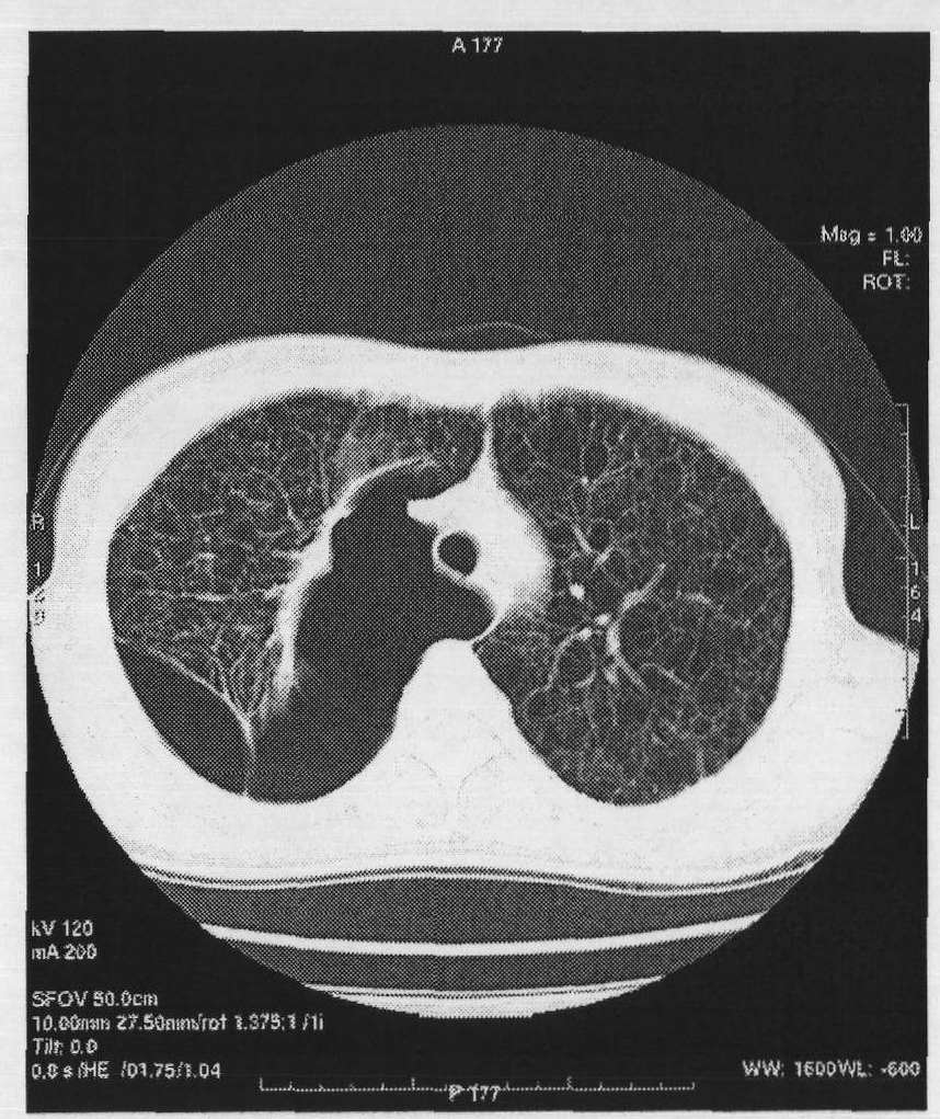
[0081] Step 1, perform a CT scan on the chest to obtain all original chest images related to the lungs, as shown in 3a;

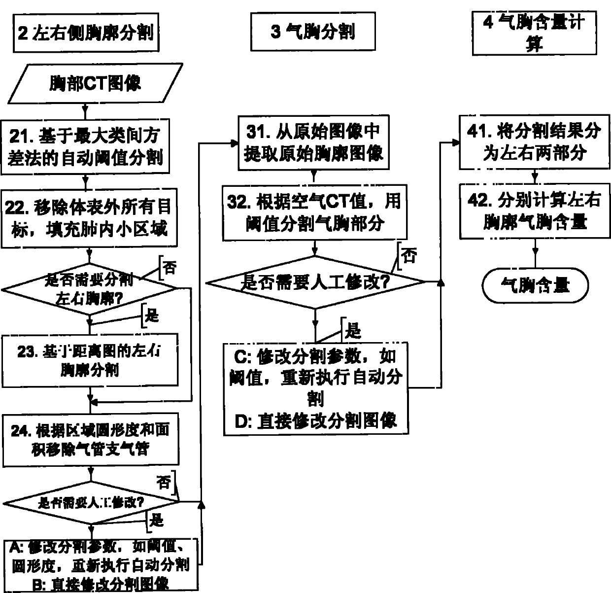
[0082] Step 2: Segment the thorax for each original thoracic image to obtain a complete separated image of the left and right sides of the thorax, such as Figure 3f shown;

[0083] Step 3, segment the pneumothorax for each original chest image, separate the pneumothorax, and obtain the image of the pneumothorax, such as Figure 5b shown;

[0084] Step 4, calculate the proportion of pneumothorax in each unilateral thorax.

[0085] Please also refer to figure 2 , figure 2 for figure 1 Further detailed flow chart of steps 2 to 4....


PUM


Abstract

The invention relates to a method and an apparatus for measuring pneumothorax. An X-ray CT (computed tomography) device for measuring pneumothorax comprises: a scanning platform, a data collection unit, an image reconstruction unit, a user operation interface for user operation, a central control unit and a pneumothorax measurement unit, wherein the pneumothorax measurement unit is used for the pneumothorax measurement of the chest image reconstructed by the image reconstruction unit and comprises a left-and-right thorax splitting unit, a pneumothorax splitting unit and a calculation unit; the left-and-right thorax splitting unit is used for splitting left and right thoraxes of the original chest image under the control of the central control unit to obtain a chest image with left and right thoraxes separated; the pneumothorax splitting unit is used for splitting the original chest image to obtain an image only with pneumothorax; and the calculation unit is used for calculating the areas of the left and right thoraxes according to the chest image with left and right thoraxes separated obtained through the splitting of the left-and-right thorax splitting unit, calculating the area of the pneumothorax according to the image only with pneumothorax obtained by the splitting of the pneumothorax splitting unit and calculating the proportion of the pneumothorax in the left and right thoraxes according to the following formula.

Description

[technical field] [0001] The invention relates to the field of medical technology, in particular to a method and a device for measuring pneumothorax based on computed tomography (CT) images. [Background technique] [0002] The lungs of the human body are firstly protected by a layer of lung pleura, which is protected by a layer of wall pleura outside the lung pleura, and the outside of the wall pleura is the thorax. In the case of a normal lung, the lung pleura and the parietal pleura fit well and move with each other. But if there is a problem with the parietal pleura of the lungs, a leak can develop that allows air to enter between the lung pleura and the parietal pleura, creating what is known as a pneumothorax. Pneumothorax (PTX) is a condition that compresses the normal exhalation and inhalation of the lungs, and when this pneumothorax reaches a certain proportion in the thorax, it becomes life-threatening. [0003] At present, there are two main methods for doctors t...

Claims


Application Information

Patent Timeline
IPC IPC(8): A61B6/03
Inventor 赵静焦洁史轶伦
Owner GE MEDICAL SYST GLOBAL TECH CO LLC
Why Eureka
Try Eureka