Method for automatic recognition and stage compression of medical image regions of interest based on artificial neural network

An artificial neural network and medical image technology, applied in biological neural network models, image analysis, image communication, etc., can solve problems such as lack of accuracy in regions of interest, large medical image files, and impact on diagnosis quality, so as to improve transmission rate, Easy to read, improve the effect of service level

Inactive Publication Date: 2012-01-25
BAILEAD TECH CO LTD
View PDF6 Cites 74 Cited by
  • Summary
  • Abstract
  • Description
  • Claims
  • Application Information

AI Technical Summary

Problems solved by technology

Existing telemedicine diagnosis systems often rely on specific hardware systems to remotely transmit patient information such as medical records and medical pictures through files or videos, often requiring doctors to conduct real-time consultations through the network, which requires high diagnostic doctors. Patient information such as transmitted medical images, because medical image files are generally large, limited by bandwidth and other factors, the transmission speed is slow, the effect is not good, and the quality of diagnosis is also affected
[0003] In the current medical image compression technology, although it is proposed to use different compression methods for regions of interest (ROI) and non-interest regions to improve transmission efficiency, the main problem is that doctors need to determine the region of interest through a manual interface, which is subject to the doctor's experience. , unit human resource conditions and subjective factors, lack of fixed standards to measure the accuracy of interest areas

Method used


Image

  • Method for automatic recognition and stage compression of medical image regions of interest based on artificial neural network
  • Method for automatic recognition and stage compression of medical image regions of interest based on artificial neural network
  • Method for automatic recognition and stage compression of medical image regions of interest based on artificial neural network

Examples


Embodiment Construction

[0029] The present invention will be described in detail below in combination with specific embodiments.

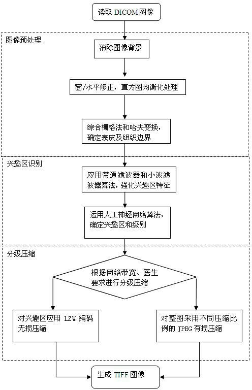
[0030] like figure 1 As shown, the method for compressing image files in the medical image transmission process of the digital diagnostic system involved in the present invention is realized by the following steps:

[0031] Step 1: Preprocessing:

[0032] Collect medical digital images on the client side, that is, DICOM images, and preprocess the image files, including background elimination, window / level correction, histogram equalization, and removal of noise and irrelevant information.

[0033] The image background includes white marks at the edge of the X-ray film due to collimator blockage, artificially annotated information, and areas outside the outline of the patient. Removing the background will help enhance the visual quality of the image while reducing the workload for subsequent processing.

[0034] In order to ensure that the diagnosis-related images are n...


PUM


Abstract

The invention relates to a method for automatic recognition and stage compression of medical image regions of interest based on an artificial neural network. Medical image files in a digital diagnostic system are generally larger, and due to the limitation by factors of bandwidth and the like, the transmission speed is low, the effect is not good, and the diagnosis quality is influenced. By the method, medical digital images are subjected to noise elimination, the tissue outline of a human body is recognized, tissue images are subjected to multiple times of overlay operation, image features of the regions of interest are strengthened, feature values are extracted, classification is performed by using an artificial neural network method, the regions of interest and corresponding levels are determined, and tagged image file format (TIFF) images are generated in different compression modes according to different levels of the regions of interest and non-regions of interest. By the method, the medical image files are greatly lessened, the transmission speed is increased, and effective necessary information used for diagnosis and treatment in the images is kept, so the method facilitates the reading of doctors, and can be applied to the digital diagnostic system and a remote medical system, and improve the diagnosis and treatment efficiency and effect.

Description

technical field [0001] The invention relates to a medical image processing method, in particular to an artificial neural network-based method for automatic identification and hierarchical compression of medical image interest areas. Background technique [0002] The digital health diagnosis system is an important part of realizing the digitization and networking of medical units. Existing telemedicine diagnosis systems often rely on specific hardware systems to remotely transmit patient information such as medical records and medical pictures through files or videos, often requiring doctors to conduct real-time consultations through the network, which requires high diagnostic doctors. Patient information such as transmitted medical images, because medical image files are generally large, limited by bandwidth and other factors, the transmission speed is slow, the effect is not good, and the quality of diagnosis is also affected. [0003] In the current medical image compress...

Claims


Application Information

Patent Timeline
Patent Type & Authority Applications(China)
IPC IPC(8): G06T7/00G06N3/02H04N7/26H04N19/167H04N19/30
Inventor 周明张雪英
Owner BAILEAD TECH CO LTD
Why Eureka
Try Eureka