Method and device for extracting skeletons of brain CT (computerized tomography) image

A CT image, brain technology, applied in the computer field, can solve problems such as errors

Inactive Publication Date: 2012-04-18
NEUSOFT CORP
View PDF2 Cites 11 Cited by
  • Summary
  • Abstract
  • Description
  • Claims
  • Application Information

AI Technical Summary

Problems solved by technology

[0008] from figure 2 It can be clearly seen in the figure that the gray value distribution range of the pixels of the two CT images that are also brain tissue is very different. If the bone threshold range is set in advance to remove the brain bone tissue, an error will occur. That is, it is possible to leave images that should be brain bones, or remove images that should not be brain bones

Method used


Image

  • Method and device for extracting skeletons of brain CT (computerized tomography) image
  • Method and device for extracting skeletons of brain CT (computerized tomography) image
  • Method and device for extracting skeletons of brain CT (computerized tomography) image

Examples


Embodiment Construction

[0055] The following will clearly and completely describe the technical solutions in the embodiments of the present invention with reference to the accompanying drawings in the embodiments of the present invention. Obviously, the described embodiments are only some, not all, embodiments of the present invention. Based on the embodiments of the present invention, all other embodiments obtained by persons of ordinary skill in the art without creative efforts fall within the protection scope of the present invention.
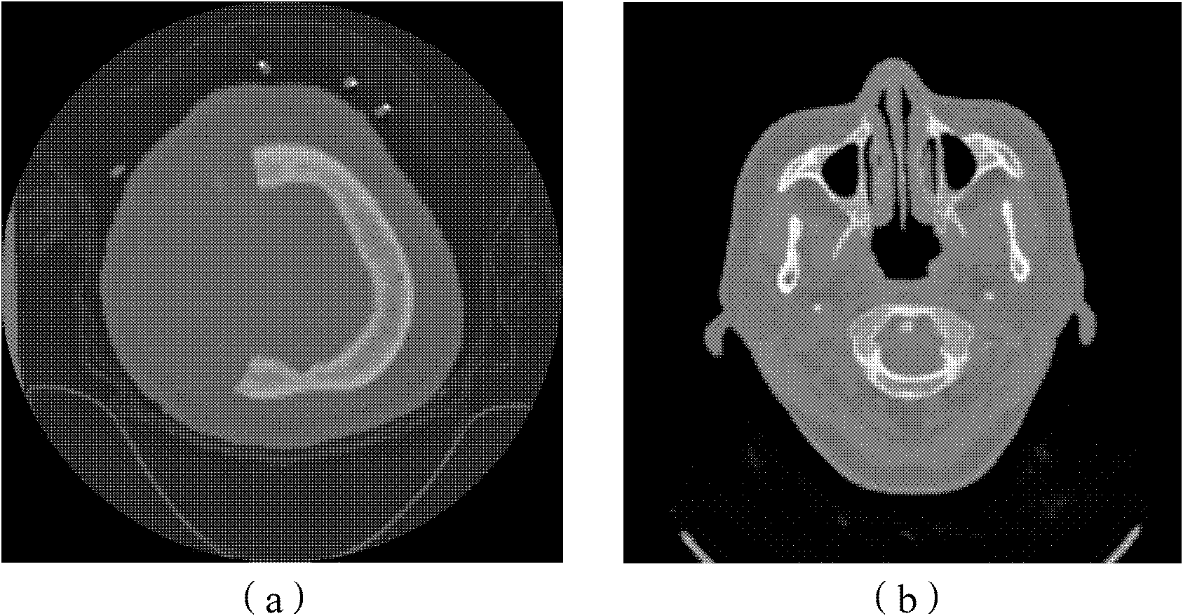
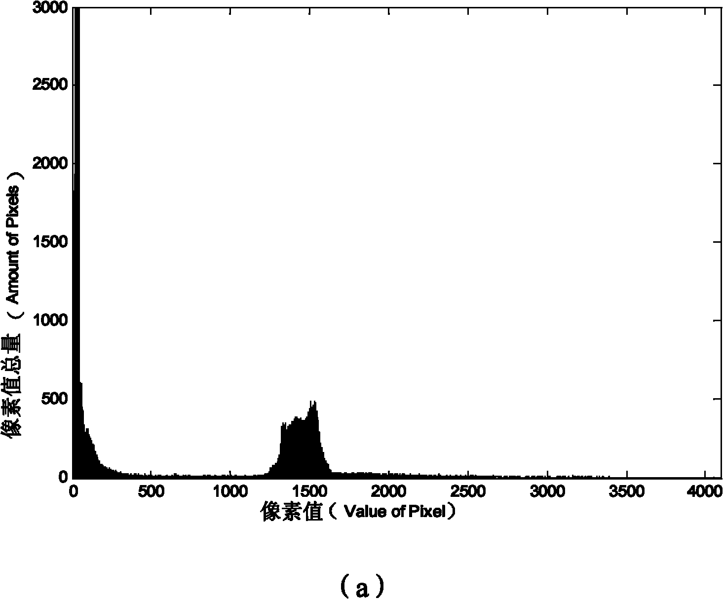
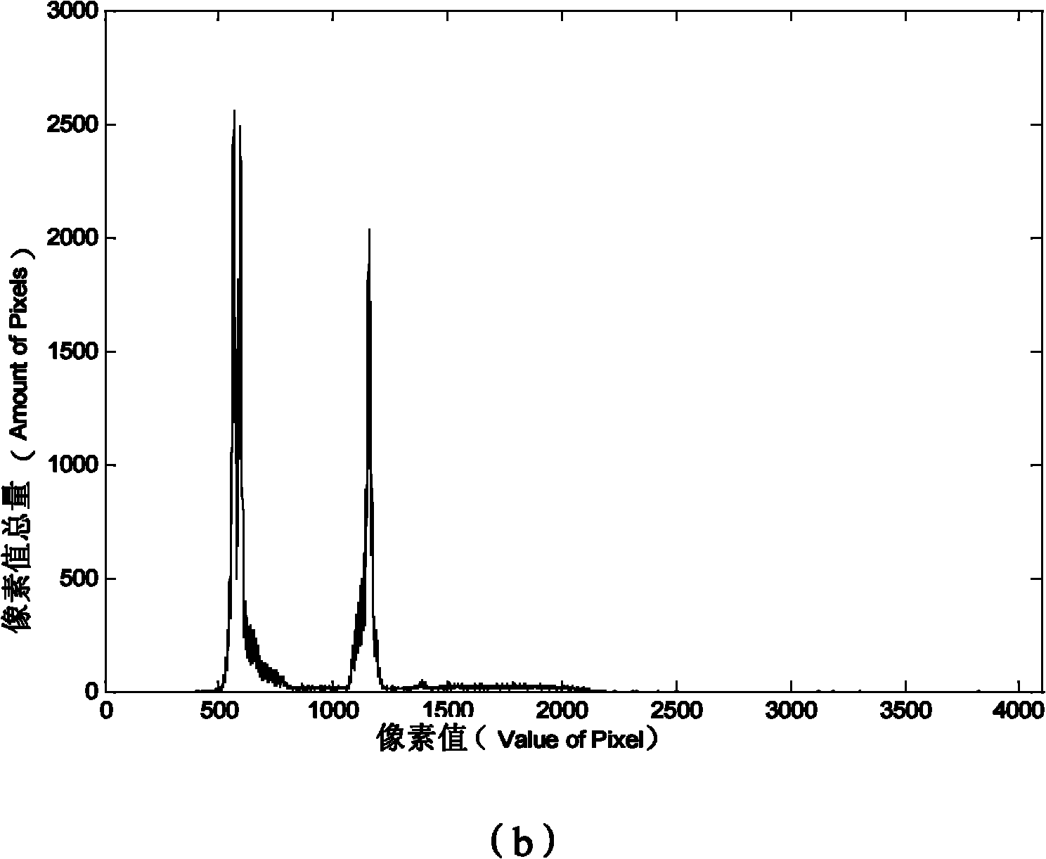
[0056] The idea of ​​this application is to remove the background pixels in the brain CT image first, and then distinguish the brain skeleton from the rest of the brain tissue on the remaining whole brain image, so as to extract the brain skeleton CT image, which will be used as a brain image for subsequent CTA images in the future. Removal of bone provides a reliable basis.

[0057] see image 3 , which is a flow chart of a method for extracting brain CT image bo...


PUM


Abstract

The application discloses a method and device for extracting skeletons of a brain CT (computerized tomography) image. The method comprises the steps of: carrying out grey mapping on pixel points of an original brain CT image to obtain a corresponding grey column diagram, and acquiring a first preset division threshold according to the grey column diagram; calculating a background pixel grey threshold according to the first preset division threshold, and removing background pixel points in the original brain CT image according to the background pixel grey threshold to obtain a pre-processed brain CT image; calculating a skeleton threshold based on the pre-processed brain CT image; and searching all pixel points of which the grey values are larger than or equal to the skeleton threshold in the pre-processed brain CT image, and extracting the image corresponding to the pixel points, wherein the image represents brain skeletons of the brain CT image. By applying the invention, the skeleton extraction error caused by carrying out empirical value setting on the skeleton threshold in advance is avoided; in addition, the method and device disclosed by the application can be used for extracting the image of brain skeletons within short time and do not bring negative influence to operation speed of integral algorithm.

Description

technical field [0001] The invention relates to the field of computer technology, in particular to a method and device for extracting bones from brain CT images. Background technique [0002] CT is a full-featured disease detection instrument, which is the abbreviation of computerized X-ray tomography. CT images are expressed in different gray scales, reflecting the degree of absorption of X-rays by organs and tissues. Therefore, like the black-and-white images shown in X-ray images, black shadows indicate low-absorption areas, that is, low-density areas, such as lungs; white shadows indicate high-absorption areas, that is, high-density areas, such as bones. Although the difference in density of human soft tissue is small, although the absorption coefficient is much closer to that of water, it can also be imaged by contrast. This is the outstanding advantage of CT. Therefore, CT can better display the organs composed of soft tissues, such as the brain, spinal cord, medias...

Claims


Application Information

Patent Timeline
Patent Type & Authority Applications(China)
IPC IPC(8): G06T7/00A61B6/03G06T7/10G06T7/136G06T7/194
Inventor 赵大哲韩芳芳覃文军杨金柱栗伟袁玉亮任福龙
Owner NEUSOFT CORP
Why Eureka
Try Eureka