

Training device for puncture operation
The technology of a training device and a puncture needle is applied in the field of medical teaching appliances, which can solve the problems of simulated venous cavity leakage and the inability to continue the puncture operation training, and achieves the effects of convenient teaching, simple implementation and improved teaching quality.
- Summary
- Abstract
- Description
- Claims
- Application Information
AI Technical Summary
Problems solved by technology
Method used
Image

Examples
Embodiment 1
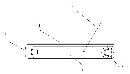
[0015] Embodiment one: if figure 1 As shown, the present invention includes a puncture model, a puncture needle 2 and a monitoring system 3. The puncture model is mainly composed of an epidermis 11 and a vein 13. The monitoring system 3 includes a monitor, a camera 31 and an illuminating lamp 32. The camera 31 and the illuminating lamp 32 are placed on In the vein 13, and are respectively electrically connected to the monitor.
[0016] The above-mentioned human body components are self-made simulation models. For the convenience of implementation, there is no need to add blood to the simulated veins 13 , but to provide accommodating space for the camera 31 and the lighting lamp 32 of the monitoring system 3 .
[0017] During operation, the puncture needle 2 enters the vein 13 from the epidermis 11, and the camera 31 can collect images of the puncture needle 2 entering the vein 13 in real time, and observe the operation process and penetration depth through the monitor, and the...
Embodiment 2
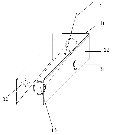
[0018] Embodiment two: if figure 2 As shown, the present invention includes a puncture model, a puncture needle 2 and a monitoring system 3. The puncture model is mainly composed of epidermis 11, muscle tissue 12 and vein 13. The monitoring system 3 includes a monitor, a camera 31 and a lighting lamp 32, and the camera 31 and lighting The lamps 32 are placed in the muscle tissue 12, and are electrically connected to monitors, respectively. The above-mentioned human body components are all self-made simulation models. For the convenience of implementation, it is further required that the simulated veins 13 and muscle tissue 12 are transparent, and the simulated epidermis 11 is opaque.
[0019] During operation, the puncture needle 2 enters the vein 13 through the epidermis 11 and muscle tissue 12 in turn, and the camera 31 can collect the images of the puncture needle 2 entering the vein 13 in real time with the help of the lighting lamp 32, and transmit them to the monitor fo...
Embodiment 3
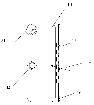
[0020] Embodiment three: as image 3 As shown, the present invention includes a puncture model, a puncture needle 2 and a monitoring system 3, the puncture model mainly consists of a pleural effusion balloon 14, ribs 15 and chest rib skin 16, and the monitoring system 3 includes a monitor, a camera 31 and an illuminating lamp 32 , the camera 31 and the lighting lamp 32 are placed in the pleural effusion balloon 14, and are electrically connected to the monitor respectively.
[0021] The above-mentioned human body components are all self-made simulation models. For the convenience of implementation, it is further required that no pleural effusion drainage fluid is added to the simulated pleural effusion balloon 14, but a placement space for the camera 31 and the lighting 32 of the monitoring system 3 is provided. .
[0022] During operation, the puncture needle 2 enters the pleural effusion balloon 14 from the entrance of the gap between the ribs 15, and the camera 31 can coll...
PUM
 Abstract
Description
Claims
Application Information