
Reduction and removal of artifacts from a three-dimensional dental X-ray data set using surface scan information

A surface scanning, X-ray technology, applied in the direction of dental radiology, dentistry, image data processing, etc., can solve the problem of manual intervention, no provision and so on

Active Publication Date: 2013-01-02
DENTAL IMAGING TECH
View PDF9 Cites 20 Cited by
  • Summary
  • Abstract
  • Description
  • Claims
  • Application Information

AI Technical Summary

Problems solved by technology

However, no details are provided on how the artifacts were removed, and the "fusion" disclosed in the '367 publication uses pre-positioning techniques that require identification of registration points on the screen or other manual means prior to merging the data
Although this pre-positioning makes the task of merging two sets of data substantially easier than a fully automated approach, this approach requires manual intervention

Method used


Image

  • Reduction and removal of artifacts from a three-dimensional dental X-ray data set using surface scan information
  • Reduction and removal of artifacts from a three-dimensional dental X-ray data set using surface scan information
  • Reduction and removal of artifacts from a three-dimensional dental X-ray data set using surface scan information

Examples


Embodiment Construction

[0024] Before any embodiments of the invention are explained in detail, it is to be understood that the invention is not limited in its application to the details of construction and the arrangement of components set forth in the following description or shown in the following drawings. The invention is capable of other embodiments and of being practiced or being carried out in various ways.

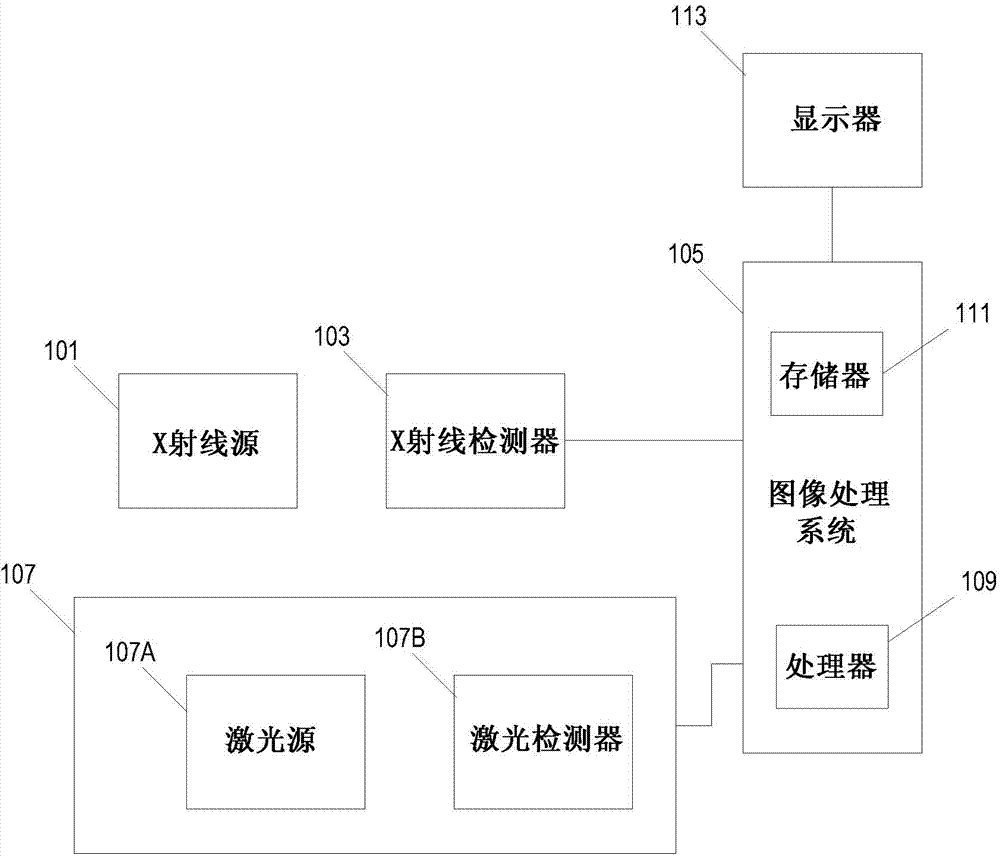
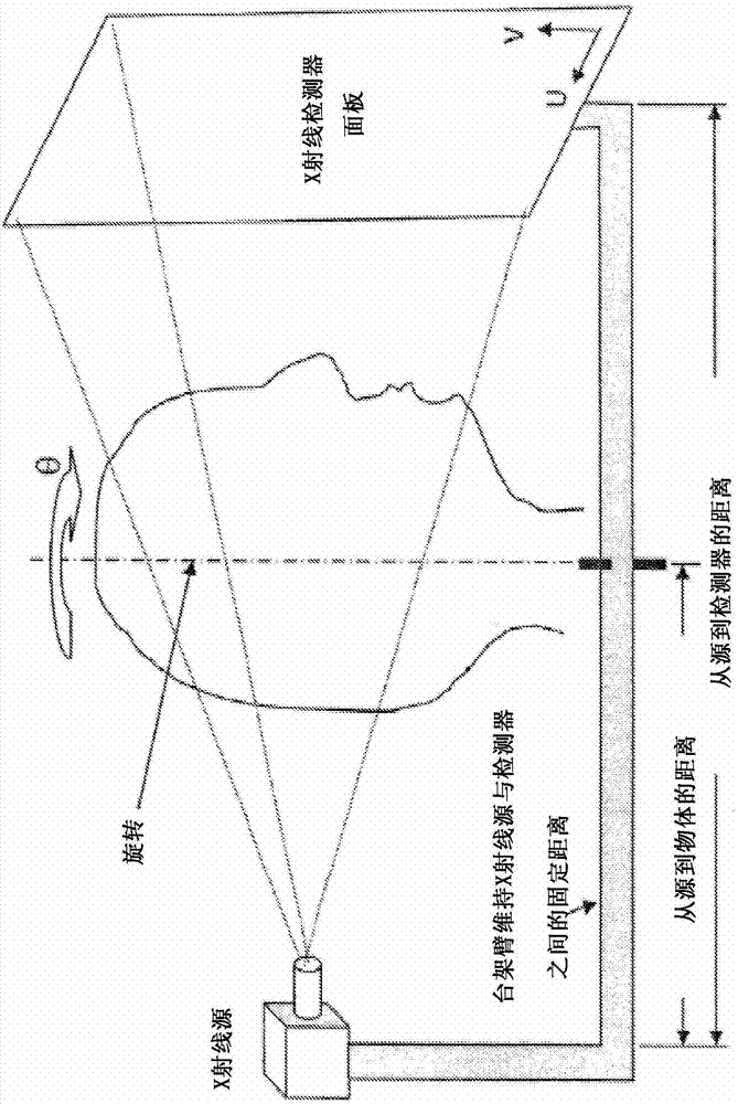
[0025] Figure 1A is a block diagram illustrating components of a system for removing artifacts from a three-dimensional digital CT model of a patient's teeth. The system can also be used to create 3D digital models of the patient's jaw and other facial bones and tissues. The system includes an X-ray source 101 and an X-ray detector 103 . An X-ray source 101 is positioned to project X-rays towards the patient's teeth. The X-ray detector 103 is placed on the opposite side of the patient's teeth - either in the patient's mouth or on the opposite side of the patient's head. The X-rays fr...


PUM


Abstract

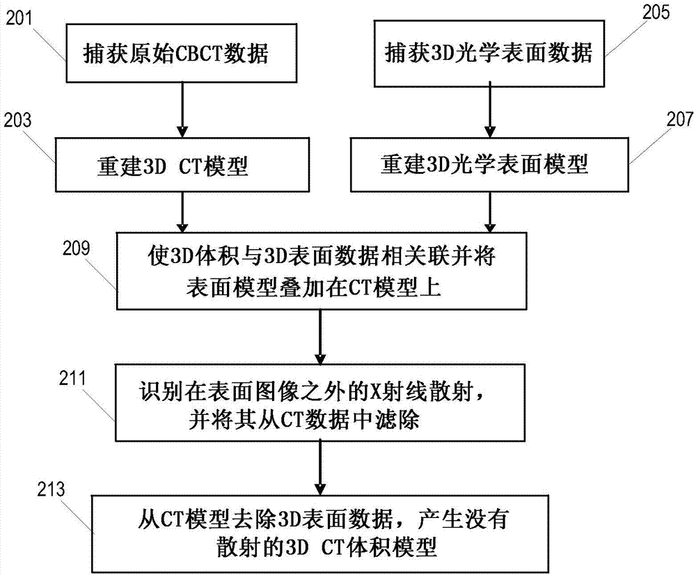
A system for removing artifacts caused by x-ray reflective materials from an x-ray image of a patient's teeth. The system includes an x-ray source, an x-ray detector that captures several x-ray images, and a surface scanner that captures a surface scan of the patient's teeth. An image processor generates three-dimensional models from the optical surface data and the CT volumetric data. The models are resized and oriented to be of the same scale and orientation and then overlaid to create a combined data set. Data points that extend beyond the surface of the patient's teeth in the surface model are identified and may be removed if it is determined that they are artifacts. An artifact-reduced CT model is then displayed.

Description

[0001] related application [0002] This patent application claims REDUCTION AND REMOVAL OF ARTIFACTS FROM A THREE-DIMENSIONALDENTAL X-RAY DATA SET USING SURFACE SCAN, filed April 20, 2010. INFORMATION), the entire contents of which are hereby incorporated by reference. technical field [0003] The present invention relates to dental imaging technology. More specifically, the invention relates to reconstructing a three-dimensional image of a patient's teeth using both x-ray and surface scanning techniques. Background technique [0004] X-ray technology can be used to produce three-dimensional digital representations of objects using computed tomography (CT). However, metals and other objects can reflect X-rays that would otherwise pass through human tissue and can be detected by X-ray detectors. Such reflections can cause unwanted artifacts in the captured data set. This effect is particularly prevalent in dental imaging, where foreign substances such as metal fillings o...

Claims


Application Information

Patent Timeline
IPC IPC(8): A61B19/00
CPCG06T2207/10081G06T19/00A61B6/5247G06T17/00A61B6/4085G06T5/005A61B6/4435G06T2207/30036A61B5/0064G06T11/008A61B6/14G06T2210/41A61B6/5258A61B6/032A61B6/51G06T5/77A61C19/04G06T7/00
Inventor 布雷德利·S·卡尔森爱德华·马兰多拉大卫·A·谢伯克乌韦·蒙德里
Owner DENTAL IMAGING TECH
Learn More