Electric impedance tomography method of brain based on layered reconstruction

A technology of electrical impedance tomography, imaging method, applied in diagnosis, diagnostic recording/measurement, medical science, etc., can solve problems such as skull layer error reflection, reconstruction method disclosure, skull artifact, etc.

Active Publication Date: 2013-03-13
FOURTH MILITARY MEDICAL UNIVERSITY
View PDF3 Cites 11 Cited by
  • Summary
  • Abstract
  • Description
  • Claims
  • Application Information

AI Technical Summary

Problems solved by technology

[0004] However, there are still two deficiencies in the prior art methods in the application of human brain imaging
First of all, the existing technology method only introduces the prior information of the skull into the forward calculation model, and does not improve the algorithm for the skull. When the target area of ​​impedance change in the cranial cavity is close to the skull area, it can be seen through the existing fusion imaging method The skull layer without impedance change will also be wrongly reflected on the impedance image, forming skull artifacts and affecting the correct judgment of the image
In addition, due to the influence of the high resistivity of the skull, the interference of the scalp area can easily overwhelm the target of impedance changes in the cranial cavity, resulting in the inability to reconstruct the target
At present, in the field of electrical impedance tomography, there is no disclosure of a reconstruction method that effectively solves the above two problems

Method used


Image

  • Electric impedance tomography method of brain based on layered reconstruction
  • Electric impedance tomography method of brain based on layered reconstruction
  • Electric impedance tomography method of brain based on layered reconstruction

Examples


Embodiment 1

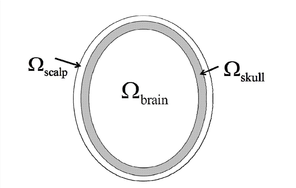
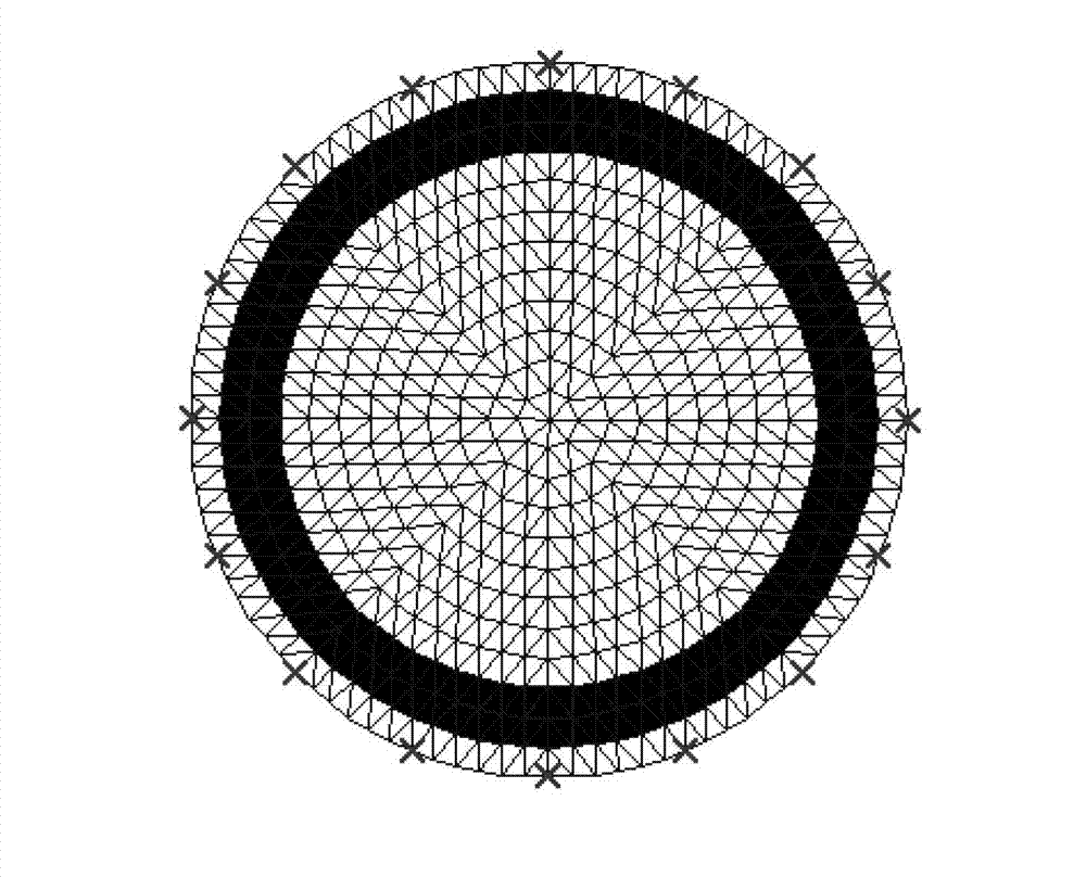
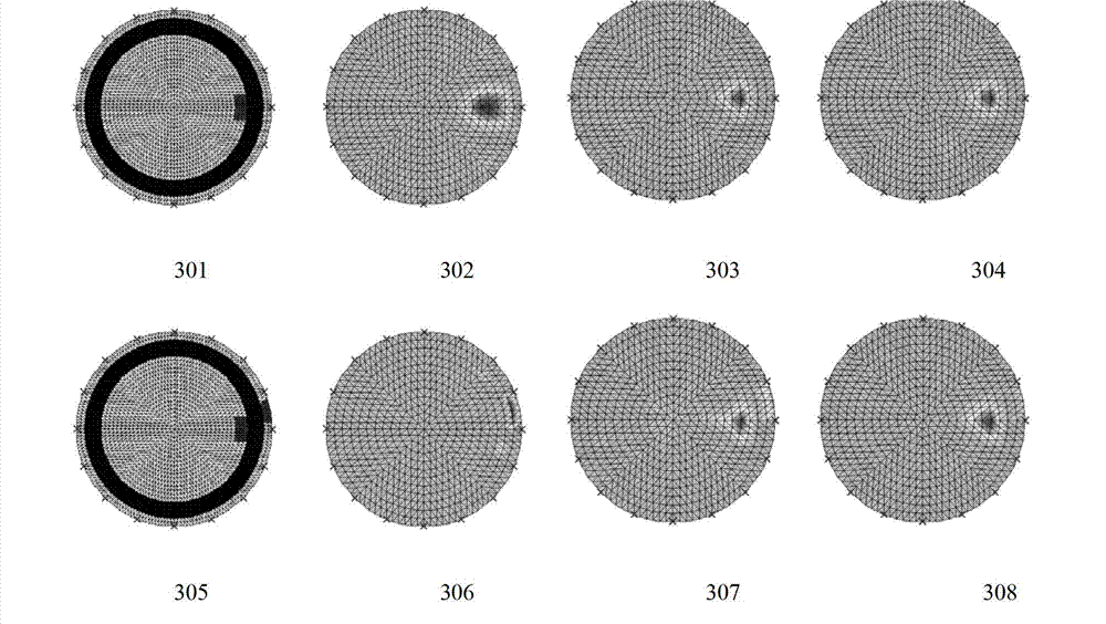
[0030] refer to figure 1 , figure 2 with image 3 , taking the computer simulation image as an example to further describe the present invention in detail. In this embodiment, firstly, the head model needs to be layered, and the layered diagram of the head model is as follows figure 1 Shown, divided into: scalp Ω scalp , skull layer Ω skull , cranial layer Ω brain . Therefore the electrical impedance vector to be reconstructed corresponding to the region Also divided into three vectors: scalp skull cranial cavity The vector form is expressed as: Δ ρ ^ = Δ ρ ^ scalp Δ ρ ^ skull ...

Embodiment 2

[0045] refer to Figure 4 , taking the human body data image as an example to further describe the present invention in detail. The method of the present invention can be used in combination with the fusion imaging method disclosed in the reference "An Electrical Impedance Tomography Method for Structural Information Fusion". In this embodiment, the measurement object is a patient undergoing burrhole drainage. Before performing electrical impedance tomography, a CT image of the patient's head has been obtained, so the regional shape of the scalp, skull, and cranial cavity in the head area is known. Prior Information. Using the methods disclosed in the references to obtain prior information on the human body structure, using the fuzzy clustering segmentation method to obtain the scalp layer, skull layer and cranial cavity layer respectively from the CT image, performing adaptive finite element segmentation on the head region, and finally collecting the electrical impedance Af...

Embodiment 3

[0048] For the case where the impedance change target in the cranial cavity is not close to the skull layer, the skull artifact can be ignored, so only the second step of hierarchical reconstruction can be used to reduce the interference of the scalp layer. Its calculation method and steps are similar to those in Example 1, just ignore the first step of layered reconstruction (that is, the constraint parameter μ is 0), and directly use the results obtained by the existing technology for the second step of layered reconstruction can be constructed.


PUM


Abstract

The invention discloses an electric impedance tomography method of a brain based on layered reconstruction, which comprises the steps as follows: layering a head model; layering the head model as a scalp layer omega scalp, a skull layer omega skull and a brain layer omega brain according to space distribution prior information; introducing a constraint matrix D to further layer and reconstruct so as to reduce artificial images of the skull layer; and layering and reconstructing the reconstructed head model in a second step to further reduce the interference of the scalp layer and highlight the target in the brain. By means of concept of layered image reconstruction, the method has the benefits of reducing the skull artificial images, lowering the interference of the scalp layer and improving the imaging quality.

Description

technical field [0001] The invention belongs to the technical field of electrical impedance tomography, and relates to a brain electrical impedance tomography method based on layered reconstruction. Background technique [0002] Electrical impedance tomography (Electrical Impedance Tomography, EIT) technology is to apply a weak alternating current excitation on the surface of the object to be tested, detect the voltage distribution of the boundary of the measured area from the corresponding detection electrodes, and reconstruct the object through the algorithm. The distribution image of electrical impedance or the distribution image of electrical impedance change inside the measured area. When the object to be measured is a living body, it can further reflect the change of the electrical impedance characteristic of the internal tissue of the living body and the change of the tissue function. EIT technology has the advantages of small imaging equipment, low cost, harmless, a...

Claims


Application Information

Patent Timeline
Patent Type & Authority Applications(China)
IPC IPC(8): A61B5/053
Inventor 董秀珍徐灿华付峰史学涛尤富生杨滨代萌
Owner FOURTH MILITARY MEDICAL UNIVERSITY
Why Eureka
Try Eureka