Method for identifying positive cells and negative cells of immunologic tissue

A positive cell, negative cell technology, applied in the medical field, can solve the problem that another batch of samples cannot be guaranteed to be effective

Active Publication Date: 2013-04-03
上海启肽智能科技有限公司
View PDF2 Cites 29 Cited by
  • Summary
  • Abstract
  • Description
  • Claims
  • Application Information

AI Technical Summary

Problems solved by technology

The problem with this method is that the judgment threshold of positive pixels is a fixed value obtained from a batch of sample images, and the segmentation result depends on the processed image. When the image has deviations in color and brightness, it cannot be guaranteed to be valid for another batch of samples. , so there is a robustness problem in the application of this method

Method used


Image

  • Method for identifying positive cells and negative cells of immunologic tissue
  • Method for identifying positive cells and negative cells of immunologic tissue
  • Method for identifying positive cells and negative cells of immunologic tissue

Examples


Embodiment Construction

[0050] The purpose of the invention of the present invention will be described in further detail below in conjunction with the accompanying drawings and specific embodiments, and the embodiments cannot be repeated here one by one, but the implementation of the present invention is not therefore limited to the following embodiments.

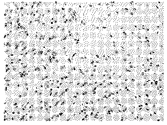
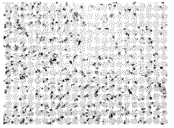
[0051] First, extract the immunohistochemical image: perform full-stained immunohistochemical staining on the tissue section, use DBA to stain the positive product, use hematoxylin to counterstain the nucleus, and obtain a digital image with a resolution of 697×1000 pixels. Figure 1 to Figure 3 The R, G, and B components of the immunohistochemical images are shown, respectively.

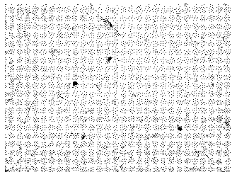
[0052] Figure 1 to Figure 3 There is a certain degree of color cast. exist Figure 4 In , the pixels satisfying R>B in RGB format are represented by white, and it can be found that there are many pixels satisfying R>B in the background part outside the cell nucleus. ...


PUM


Abstract

The invention discloses a method for identifying positive cells and negative cells of an immunologic tissue. The method provided by the invention can be used for determining a corresponding threshold value without using a certain threshold in a self-adaptive manner according to an own maximum between-cluster variance method of immunohistochemical image data to be processed, and can be adapted to deviations of an image on the color and the light intensity, so that the method is more stable when being compared with the conventional method; when nucleuses are judged to be positive or negative, dyeing processing information of the immunohistochemical image is used as prior information without manual marking, so that the intervene of subjective factors can be avoided, the full-automatic processing can be realized, and the stability of analysis efficiency and process quality can be improved.

Description

technical field [0001] The invention relates to the field of medical technology, in particular to a method for identifying immune tissue positive cells and negative cells. Background technique [0002] Immunohistochemical technology combines fluorescent or colorable chemical substances on antibodies, and uses the specific binding reaction between antigens and antibodies in immunological principles to detect the presence of target antigens in cells or tissues. important method of diagnosis. The color images obtained by imaging cells or tissues under a microscope after immunohistochemical staining, because the staining process involves multiple steps and factors, the staining results will have different degrees of difference in the overall brightness and the color of positive substances, lacking consistency and repeatability. The method of artificially marking or obtaining recognition rules based on a certain image sample is greatly affected by the subjective experience of t...

Claims


Application Information

Patent Timeline
Patent Type & Authority Applications(China)
IPC IPC(8): G06K9/00G06K9/46
Inventor 李乡儒杨坦
Owner 上海启肽智能科技有限公司
Why Eureka
Try Eureka