Detecting system and detecting method for ultrasonic blood vessel boundaries

A technology of boundary detection and blood vessels, applied in the field of medical image processing systems, can solve the problems of time-consuming, speckle noise, low resolution of ultrasonic images, etc., and achieve the effect of accurate coordinates

Active Publication Date: 2013-05-08
珠海中科先进科技产业有限公司
View PDF2 Cites 13 Cited by
  • Summary
  • Abstract
  • Description
  • Claims
  • Application Information

AI Technical Summary

Problems solved by technology

Since the intravascular ultrasound images taken for the same patient often contain thousands of images, the manual marking method is not only time-consuming, but also has poor repeatability, and is easily affected by experience and subjective factors
[0004] In addition, the existing methods of using image processing to detect the vascular lumen boundary and the media-adventitia boundary have shortcomings such as low resolution of ultrasound images, accompanied by severe speckle noise and calcification artifacts, making detection of the vascular lumen boundary and the media-adventitia boundary difficult. There are great difficulties

Method used


Image

  • Detecting system and detecting method for ultrasonic blood vessel boundaries
  • Detecting system and detecting method for ultrasonic blood vessel boundaries
  • Detecting system and detecting method for ultrasonic blood vessel boundaries

Examples


Embodiment Construction

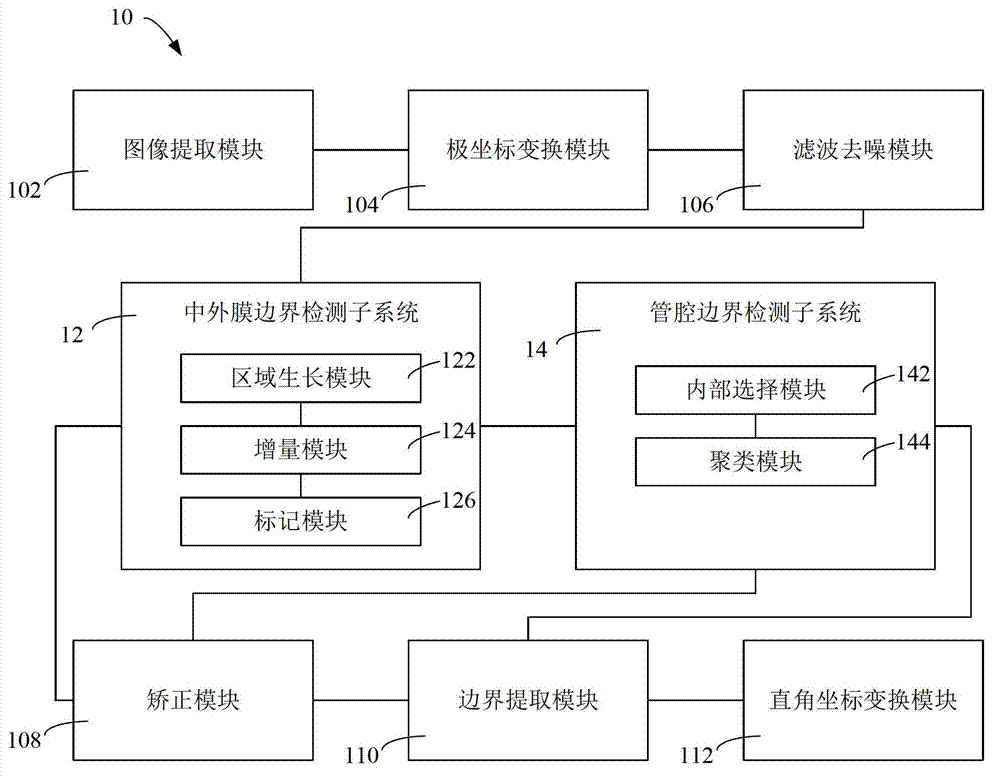
[0065] figure 1 Shown is a module structure diagram of an embodiment of the ultrasonic blood vessel boundary detection system of the present invention. According to this embodiment, the ultrasonic blood vessel boundary detection system 10 includes an image extraction module 102, a polar coordinate transformation module 104, a filtering and denoising module 106, a media-adventitia boundary detection subsystem 12 and a lumen boundary detection subsystem 14, a correction module 108, a boundary An extraction module 110 and a Cartesian coordinate transformation module 112 .

[0066] The image extraction module 102 is used to extract the characteristic image area of ​​blood vessels from the ultrasound image. The blood vessel feature image region generally has image features that are quite different from the background image, so the extraction method of the image extraction module 102 may include methods such as histogram, Fourier transform, and least squares, but is not limited the...


PUM


Abstract

A detecting system for ultrasonic blood vessel boundaries comprises an image extraction module, a tunica media and tunica adventitia boundary detecting subsystem, and a lumen boundary detecting subsystem, wherein the image extraction module is used for extracting blood vessel feature images from ultrasonic images, the tunica media and tunica adventitia boundary detecting subsystem is used for detecting the boundaries of blood vessel tunica media and blood vessel tunica adventitia from the blood vessel feature images through a region growing method, and the lumen boundary detecting subsystem is used for further detecting the blood vessel lumen boundaries in the inner regions, detected by the tunica media and tunica adventitia boundary detecting subsystem, of the boundaries of the tunica media and the tunica adventitia through a clustering method. The invention further provides a corresponding detecting method for the ultrasonic blood vessel boundaries. According to the detecting system for the ultrasonic blood vessel boundaries and the detecting method for the ultrasonic blood vessel boundaries, the images inside ultrasonic blood vessels are detected through the region growing method, so that images on two sides of each boundary of the blood vessel tunica media and tunica adventitia are easy to tell and images on two sides of each lumen boundary of the blood vessel images are easy to tell through the clustering method, and therefore detected coordinates of the boundaries of the tunica media and the tunica adventitia and the lumen boundaries are accurate.

Description

technical field [0001] The present invention relates to a medical image processing system and method, in particular, the present invention relates to a system and method for detecting the boundary of the lumen of a blood vessel and the boundary of the media and adventitia based on an ultrasonic image. Background technique [0002] Cardiovascular and cerebrovascular diseases have become the number one killer of human health. Atherosclerosis and its complications are common causes of cardiovascular and cerebrovascular diseases. The size and shape of the vessel lumen boundary and media-adventitia boundary are important indicators for measuring the degree of atherosclerosis in clinical diagnosis. [0003] In order to conveniently obtain the position information of the vascular lumen boundary and the junction of media and adventitia, blood vessel images are often taken by ultrasonography. In the existing method, manually mark the vascular lumen boundary and the media-adventitia...

Claims


Application Information

Patent Timeline
Patent Type & Authority Applications(China)
IPC IPC(8): G06T7/00G06T5/00
Inventor 杨平高智凡张元亭
Owner 珠海中科先进科技产业有限公司
Why Eureka
Try Eureka