
A kind of ultrasonic imaging device and method

An ultrasonic imaging and imaging technology, which is applied in measuring devices, ultrasonic/acoustic/infrasonic diagnosis, acoustic wave diagnosis, etc., can solve the problem that the imaging method has not reached the optimal state, and achieve the effect of increasing diagnostic confidence

Active Publication Date: 2016-05-25
SHENZHEN MINDRAY BIO MEDICAL ELECTRONICS CO LTD +1
View PDF12 Cites 1 Cited by
  • Summary
  • Abstract
  • Description
  • Claims
  • Application Information

AI Technical Summary

Problems solved by technology

However, from the perspective of actual imaging effects, conventional imaging methods are far from optimal for imaging specific areas.

Method used


Image

  • A kind of ultrasonic imaging device and method
  • A kind of ultrasonic imaging device and method
  • A kind of ultrasonic imaging device and method

Examples


Embodiment approach

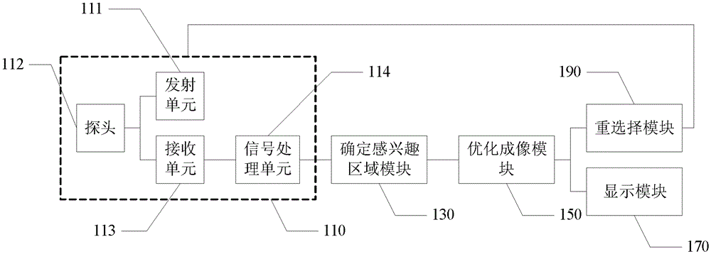
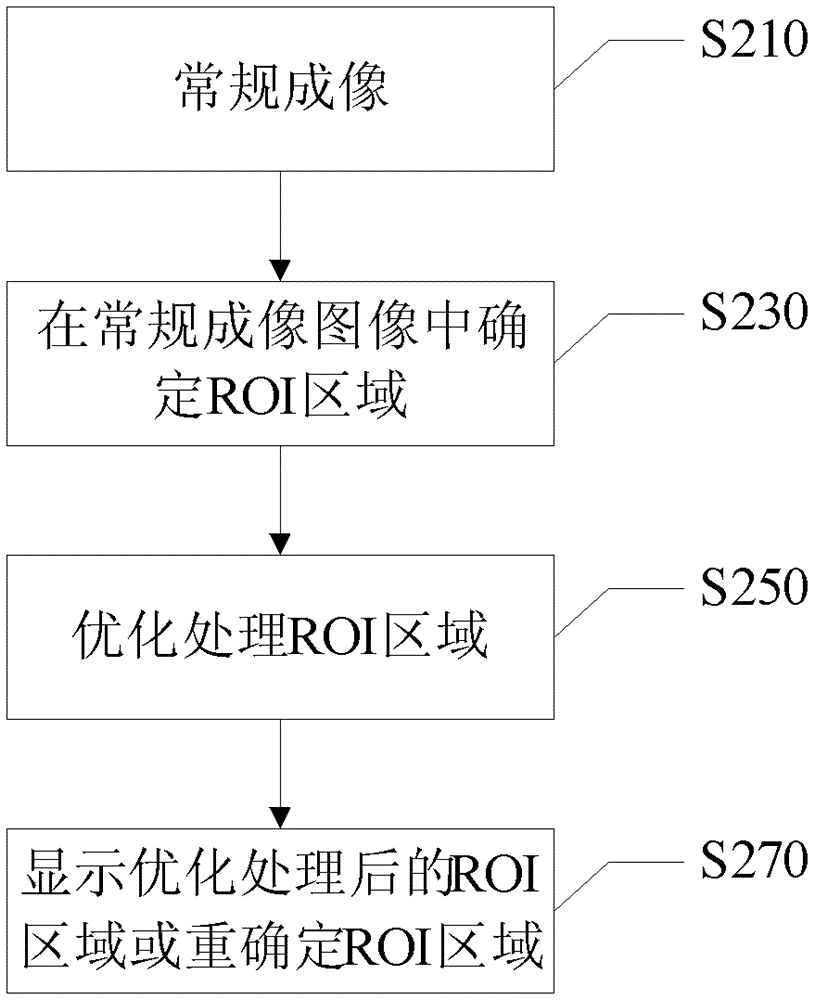
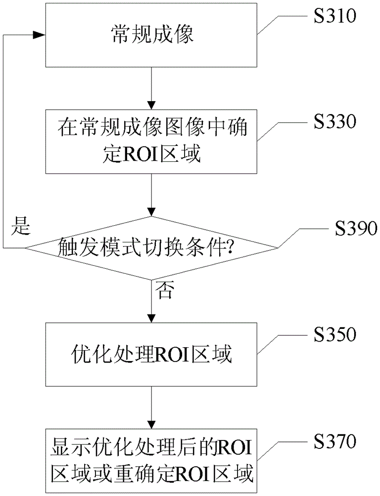
[0025] figure 1 Shown is an implementation of the ultrasonic imaging device of the present invention, including: a conventional imaging module 110 , a module for determining a region of interest 130 , and an optimized imaging module 150 . Wherein, the conventional imaging module 110 is used to transmit ultrasonic pulses to the target to be detected, receive the ultrasonic echo signal reflected by the target to be detected, and output the conventional imaging image after processing the received ultrasonic echo signal; the conventional imaging module 100 includes a transmitting unit 111, a probe 112, a receiving unit 113, a signal processing unit 114, etc. That is to say, the conventional imaging module 110 can be an ultrasound imaging module well known to those skilled in the art, wherein the transmitting unit 111 transmits ultrasonic waves into the human body through the probe 112. After being reflected by the tissue of the human body, it is received by the receiving unit 113 ...


PUM


Abstract

The invention discloses an ultrasonic imaging device and method, wherein the device includes: a conventional imaging module, which is used to transmit ultrasonic pulses to a target to be detected, receive ultrasonic echo signals reflected by the target to be detected, and analyze the received ultrasonic echo signals After signal processing, the conventional imaging image is output; the module of determining the region of interest is used to determine the region of interest in the conventional imaging image; the optimization imaging module is used to freeze the image region outside the region of interest in the conventional imaging image, and The optimized imaging process is performed on the region of interest, and the image of the region of interest after the optimized imaging process is output. The present invention determines the region of interest on the basis of conventional ultrasound imaging, keeps the region outside the region of interest in an image freeze state, and only optimizes the region of interest, so that each region of interest can achieve the best imaging effect, thereby It complements the deficiency of conventional imaging images and helps to increase the user's diagnostic confidence.

Description

technical field [0001] The invention relates to the field of ultrasonic imaging, in particular to an ultrasonic imaging device and method. Background technique [0002] In the conventional two-dimensional grayscale imaging method, in order to take into account the overall image performance, the ultrasound imaging frequency must meet the entire imaging depth. When the imaging depth is deep (that is, far-field imaging), the imaging frequency must be low enough to obtain sufficient penetration; however, for near-field and mid-field imaging, if still using a lower imaging frequency, the Spatial resolution will suffer. Due to the consideration of the overall image, the existing imaging methods can only be compromised. [0003] Under the same emission parameters (including emission aperture, scanning range, emission waveform, etc.), the image signal-to-noise ratio is proportional to the emission voltage of the ultrasonic probe; when the instantaneous value of the sound pressure ...

Claims


Application Information

Patent Timeline
Patent Type & Authority Patents(China)
IPC IPC(8): A61B8/00
CPCG01S7/52063G01S15/8979G01S15/8995
Inventor 史志伟
Owner SHENZHEN MINDRAY BIO MEDICAL ELECTRONICS CO LTD
Learn More