pet‑ct system with single detector

A technology of detectors and optical detectors, applied in instruments, measuring devices, scientific instruments, etc., can solve problems such as registration errors, and achieve the effect of reducing costs and simplifying registration

Active Publication Date: 2017-02-22
KONINK PHILIPS ELECTRONICS NV
View PDF8 Cites 0 Cited by
  • Summary
  • Abstract
  • Description
  • Claims
  • Application Information

AI Technical Summary

Problems solved by technology

Thus, even small amounts of misalignment in patient positioning can lead to significant registration errors in the combined or fused images

Method used


Image

  • pet‑ct system with single detector
  • pet‑ct system with single detector
  • pet‑ct system with single detector

Examples


Embodiment Construction

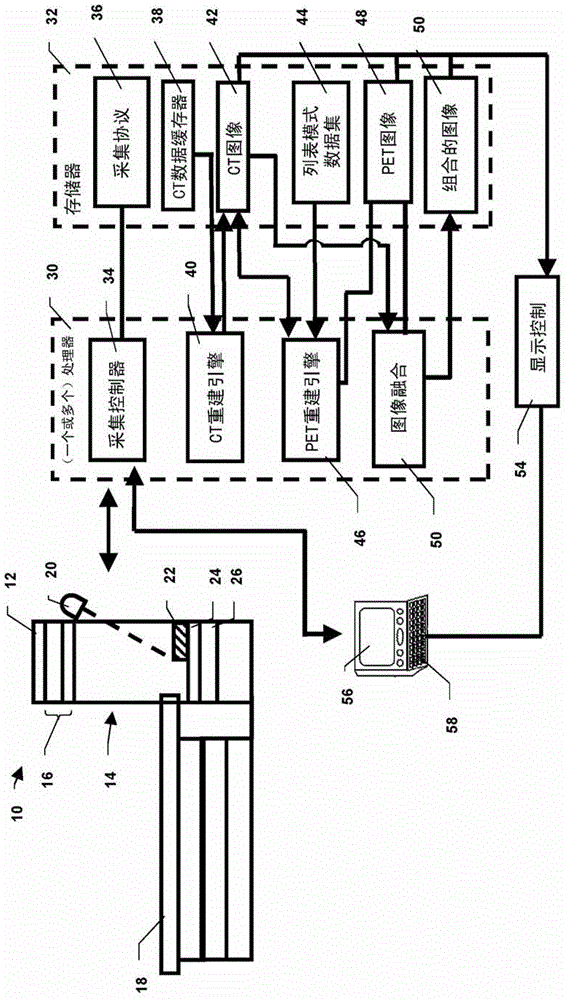
[0021] refer to figure 1 , the hybrid imaging system 10 includes a single gantry 12 that defines an examination region 14 therein. A ring of radiation detectors is disposed around the examination area, said ring of radiation detectors detecting radiation emitted by a patient or other object on the patient support 18 or passing through the patient support 18 as the patient support 18 extends into the examination area radiation on patients or other objects. exist figure 1 In an embodiment of the present invention, a transmitted radiation source 20 such as an X-ray tube and an anti-scatter grid (also called a scatter reject collimator) 22 are arranged to rotate around the examination region 14 . In one embodiment, the anti-scatter grid is movable from the examination region to acquire functional or emission data (eg, nuclear data) during a PET, SPECT, or other nuclear scan. The anti-scatter grid is a set of vanes, each of which is aligned with the focal point of the x-ray sour...


PUM


Abstract

A radiation detector (16) with a first detector layer (24) and a second detector layer (26) surrounds an examination region (14). The first layer of detectors includes a scintillator (72) and a photodetector (74) such as an avalanche photodiode. The detectors of the second detector layer (26) include a scintillator (62) and a photodetector (64). The scintillators (72) of the first layer have a smaller cross-section than the scintillators (62) of the second layer. A set, eg nine, of said first layer scintillators ( 72 ) covers each second set of scintillators ( 62 ). In CT mode, the first layer of detectors detects transmitted radiation to generate CT images with relatively high resolution, and the second layer of detectors detects PET or SPECT radiation to generate Nuclear data for rate emission images. Because the first and second layer detectors are aligned, the transmission image and the emission image are inherently aligned.

Description

technical field [0001] The following relates to the medical field, the field of medical imaging, the field of medical diagnostics, the field of positron emission tomography (PET) imaging, the field of computed tomography (CT) and related fields. Background technique [0002] The use of positron emission tomography (PET), single photon emission computed tomography (SPECT), and other imaging modalities in tumor diagnosis, assessment, and treatment planning is increasing. PET and SPECT entail administering a radiopharmaceutical to a subject (eg, a human or animal subject) and detecting radiation emitted from the subject by the radiopharmaceutical. The radiopharmaceuticals can be tailored to preferentially collect in the bloodstream or other anatomical regions of interest, thereby providing image contrast for those regions. For oncology, PET and SPECT are considered complementary to transmission computed tomography (CT) or magnetic resonance (MR), since PET and SPECT are biased...

Claims


Application Information

Patent Timeline
Patent Type & Authority Patents(China)
IPC IPC(8): A61B6/03G01T1/164
CPCA61B6/037A61B6/4028A61B6/4233A61B6/4291A61B6/4417A61B6/5229G01T1/1611G01T1/1612G01T1/1614G01T1/248A61B6/032G01T1/20181
Inventor C·赫尔曼
Owner KONINK PHILIPS ELECTRONICS NV
Why Eureka
Try Eureka