High-frequency ultrasonic superficial organ imaging method capable of lowering random noise

A technology of random noise and imaging method, applied in ultrasonic/sonic/infrasonic diagnosis, sonic diagnosis, infrasonic diagnosis, etc., can solve problems such as application limitations

Inactive Publication Date: 2013-09-18
INST OF BIOMEDICAL ENG CHINESE ACAD OF MEDICAL SCI
View PDF4 Cites 6 Cited by
  • Summary
  • Abstract
  • Description
  • Claims
  • Application Information

AI Technical Summary

Problems solved by technology

However, it is difficult for the operator to stabilize the probe at this precision, so ghosting is inevitable, which limits the application of this technology in high-frequency ultrasound imaging

Method used


Image

  • High-frequency ultrasonic superficial organ imaging method capable of lowering random noise
  • High-frequency ultrasonic superficial organ imaging method capable of lowering random noise
  • High-frequency ultrasonic superficial organ imaging method capable of lowering random noise

Examples


Embodiment

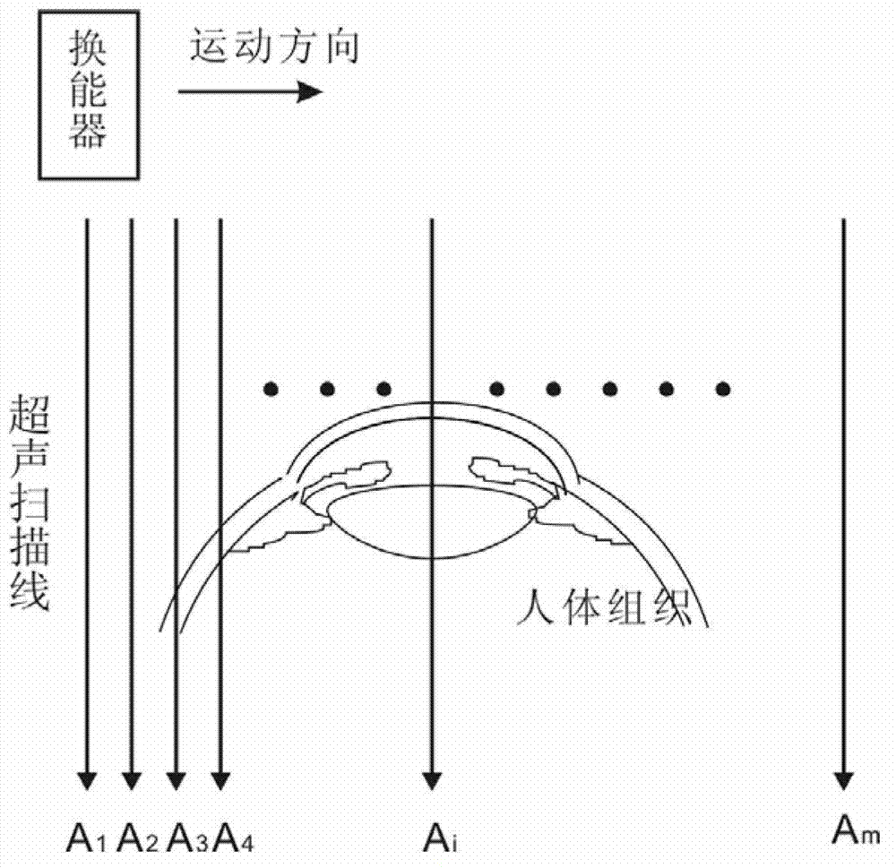
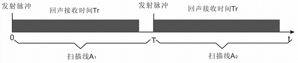
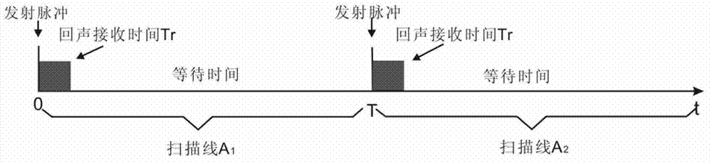
[0025] The number of scanning lines is m=500, the dwell time of each scanning line is T=200μs, the number of ultrasonic pulses transmitted at equal intervals within the dwell time T is N=4, the echo receiving time is Tr=23μs, and the time for acquiring an image is 100ms. Figure 6It is a schematic circuit diagram of line average method processing in the embodiment, including echo information receiving channel, buffer selection counter, 1-4 selection switch, buffer 1-4, adder, line memory and address pointer counter. The mutual position or connection relationship between them is: the echo information receiving channel outputs 8-bit echo information that has been transformed into a digital signal, and its output is connected to the data input terminals of the buffer 1 ~ buffer 4, and the buffer selection counter controls 1-4 The selector switch selects and sends the input clock CKin to the buffers 1 to 4 sequentially during the 4 transmission cycles within the time T=200, and the...


PUM


Abstract

The invention discloses a high-frequency ultrasonic superficial organ imaging method capable of lowering random noise. The method is characterized in that an ultrasonic transducer continuously transmits N times of ultrasonic pulses at intervals within a dwell time of 200 microseconds on each scanning position in a scanning area, after the superimposition average operation processing is carried out by adopting a linear average method for the ultrasonic echo information, and the processing result is used as a scanning line of the scanning position; and then the scanning position is moved to repeat the process until all scanning lines are completed, and the two-dimensional high-frequency ultrasonic imaging can be realized. The echo information acquired through N times of detection is superimposed and averaged by adopting the linear average method, the random noise in the echo information of the scanning line is lowered, the signal-to-noise can be theoretically improved by N times, no frame frequency is reduced, and the imaging real-time property is not influenced. Since the N-time sampling time anticipating in the average calculation is about 200 microseconds, a probe and a tissue can be easily maintained relatively still, and the ghosting phenomenon caused by the frame average technology can be avoided.

Description

technical field [0001] The invention relates to an ultrasonic imaging method, in particular to a high-frequency ultrasonic superficial organ imaging method capable of reducing random noise. Background technique [0002] The principle of ultrasonic imaging is that the ultrasonic transducer transmits ultrasonic pulses to the human tissue, and then receives the echo reflected by the human tissue, so as to obtain the one-dimensional information of the human tissue on the ultrasonic propagation channel, translate or rotate the ultrasonic emission direction, and at the same time Continuously emitting ultrasound and receiving echoes, the tissue information of the plane area scanned by the sound beam will be obtained (see figure 1 ), and reconstruct these information to obtain a two-dimensional section image of human tissue, which is commonly referred to as a B-ultrasound image. [0003] The echo information is very weak, so it needs to be amplified many times before it can be obse...

Claims


Application Information

Patent Timeline
Patent Type & Authority Applications(China)
IPC IPC(8): A61B8/00
Inventor 杨军宋学东计建军王延群
Owner INST OF BIOMEDICAL ENG CHINESE ACAD OF MEDICAL SCI
Why Eureka
Try Eureka