Magnetic resonance imaging apparatus

一种磁共振成像、图像的技术,应用在测量装置、测量磁变量、医药科学等方向,能够解决磁化并未充分恢复的状态下等问题

Inactive Publication Date: 2013-12-25
TOSHIBA MEDICAL SYST CORP
View PDF7 Cites 4 Cited by
  • Summary
  • Abstract
  • Description
  • Claims
  • Application Information

AI Technical Summary

Problems solved by technology

When this non-contrast MRA is performed using biological information (such as respiratory cycle), there is a problem that, for example, when the patient's respiratory cycle becomes extremely short, imaging is performed without sufficient magnetization recovery.

Method used


Image

  • Magnetic resonance imaging apparatus
  • Magnetic resonance imaging apparatus
  • Magnetic resonance imaging apparatus

Examples


no. 2 Embodiment approach

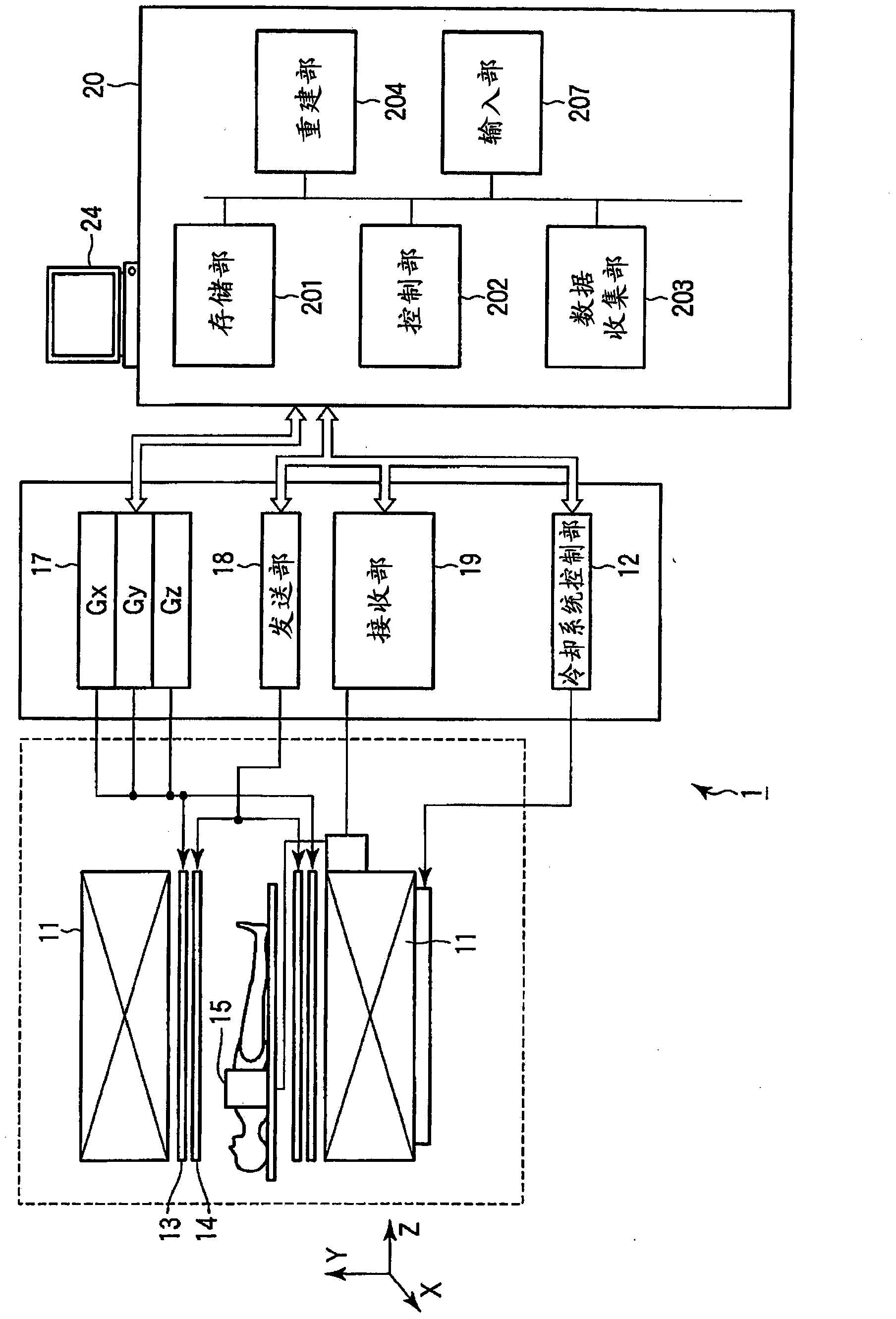
[0087] Next, a magnetic resonance imaging apparatus according to a second embodiment of the present invention will be described. The magnetic resonance imaging apparatus according to the present embodiment is an apparatus that applies temporal adjustment processing to non-contrast MRA imaging. That is, in the magnetic resonance imaging apparatus according to this embodiment, at least one of the TI, the flip angle of the IR pulse, and the flip angle of the RF pulse for imaging is adjusted according to fluctuations in biosignals such as the respiratory cycle to obtain In non-contrast MRA, which is less affected by physical activity, the above-mentioned TI is the period from when the inversion recovery pulse (IR pulse) for tagging is applied to when the first RF pulse for imaging is applied . Such a non-contrast MRA imaging method is sometimes called Time-SLIP (Time-Spatial Labeling Inversion pulse: real-time-space marker inversion pulse) method.

[0088] The so-called "additi...

Deformed example 1

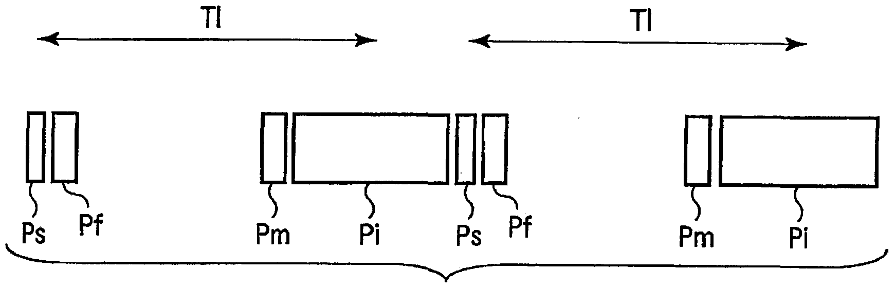
[0110] In the second embodiment, a sequence of imaging using the three-dimensional SSFP (Steady-State Free Precession: Steady-State Free Precession) method is shown (see Figure 11 , Figure 12 ). However, the present invention is not limited to this example. For example, other methods such as a three-dimensional FSE (Fast Spin-Echo) method and a three-dimensional FASE (Fast Advanced Spin-Echo) method may be used. Scan sequence for imaging. Moreover, the image collection method may be any one of single-shot or multiple-shot. if e.g. figure 2 An example is a sequence according to the single-shot 3D SSFP method, the first slice encoding is performed in imaging I, and then the second slice encoding is performed in imaging II. On the other hand, if for example Figure 11 , Figure 12 The example is based on the sequence of the three-dimensional SSFP method of multiple imaging (two imaging), the first imaging of the first slice encoding is performed in imaging I, and then th...

Deformed example 2

[0112] In the second embodiment, the case where the biological signal is set as a respiratory cycle was exemplified. However, without being limited to this example, the technical idea of ​​the present invention is also applicable to, for example, non-contrast MRA imaging using ECG waveforms or pulsation waveforms as biosignals.

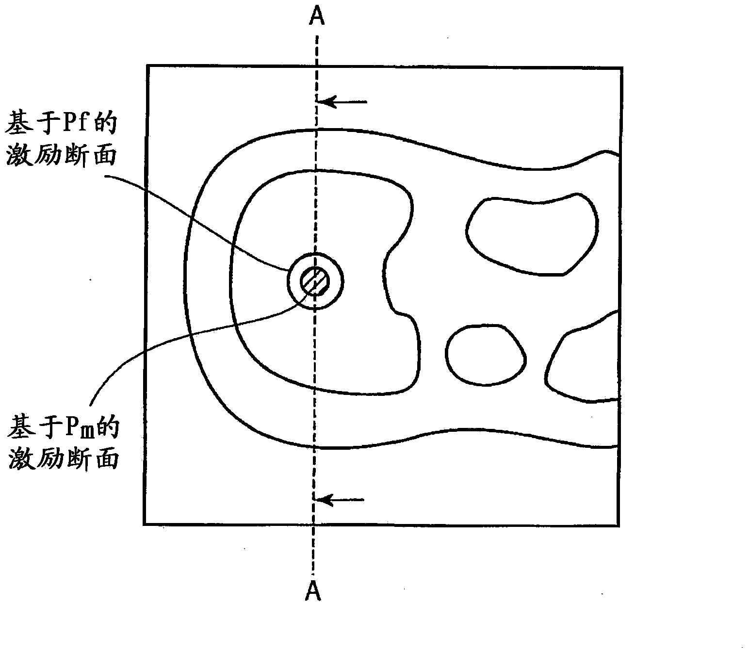
[0113] According to the configuration as described above, when non-contrast MRA imaging is performed, the fluctuation amount of the respiratory cycle is measured, and the waiting time from the tagged pulse to the imaging pulse and the inversion angle of the tagged pulse are determined according to the fluctuation amount. The suppression of the imaging pulse and the control of the flip angle of the imaging pulse make the phase of the dynamic imaging relatively correspond to the cardiac time, and the imaging conditions can be adjusted according to the mode of the respiratory cycle change. Therefore, it is possible to correct a positional shift in the dy...


PUM


Abstract

Disclosed is a magnetic resonance imaging apparatus. During myocardium perfusion imaging, the variation of position in the dynamic time-phase direction due to a respiratory body motion of the subject is detected, and the slice excitation position and the data collection position are made to follow up the body motion according to the variation. For the follow-up, a saturation pulse of space nonselection and a local selection pulse for flip back of the flip angle attributed to the saturation pulse of space nonselection are applied to the region to which a probe pulse for detecting the body motion of the subject before the heart is imaged is applied before the probe pulse is applied, thereby measuring the body motion.

Description

[0001] This application is an application for an invention patent with the application number 200980134450.7 (PCT / JP2009 / 065526), ​​the application date is September 4, 2009 (the submission date is March 4, 2011), and the invention name is "magnetic resonance imaging device". Divisional application. technical field [0002] The present invention relates to a magnetic resonance imaging apparatus that can perform high-precision myocardial perfusion imaging and non-contrast MRA (magnetic resonance angiography: magnetic resonance angiography), and can improve the diagnostic performance of the heart. Background technique [0003] A magnetic resonance imaging device is a device that, when a core group having an intrinsic magnetic moment is placed in a uniform static magnetic field, utilizes a phenomenon in which energy of a high-frequency magnetic field rotating at a specific frequency is absorbed resonantly, and the chemical and Physical microscopic information imaging, or observ...

Claims


Application Information

Patent Timeline
Patent Type & Authority Applications(China)
IPC IPC(8): A61B5/055
CPCA61B5/055A61B5/7207G01R33/4828G01R33/4838G01R33/5607G01R33/5613G01R33/56333G01R33/56366G01R33/56509G01R33/5673G01R33/5676A61B5/7285
Inventor 葛西由守
Owner TOSHIBA MEDICAL SYST CORP
Why Eureka
Try Eureka