
MR-angiography with non-cartesian signal acquisition

An angiography, non-Cartesian technique, applied to the measurement of magnetic variables, medical science, measurement devices, etc., can solve the problem of inability to obtain efficiency

Active Publication Date: 2013-01-02
SIEMENS HEALTHCARE GMBH
View PDF7 Cites 4 Cited by
  • Summary
  • Abstract
  • Description
  • Claims
  • Application Information

AI Technical Summary

Problems solved by technology

But the problem here is that no efficiency can be gained here because the time used to encode the 3D partition is the same as the time used to get the 2D layer
So in order to improve a resolution of eg 3mm in the z direction to 0.75mm a measurement time 4 times longer has to be used

Method used


Image

  • MR-angiography with non-cartesian signal acquisition
  • MR-angiography with non-cartesian signal acquisition
  • MR-angiography with non-cartesian signal acquisition

Examples


Embodiment Construction

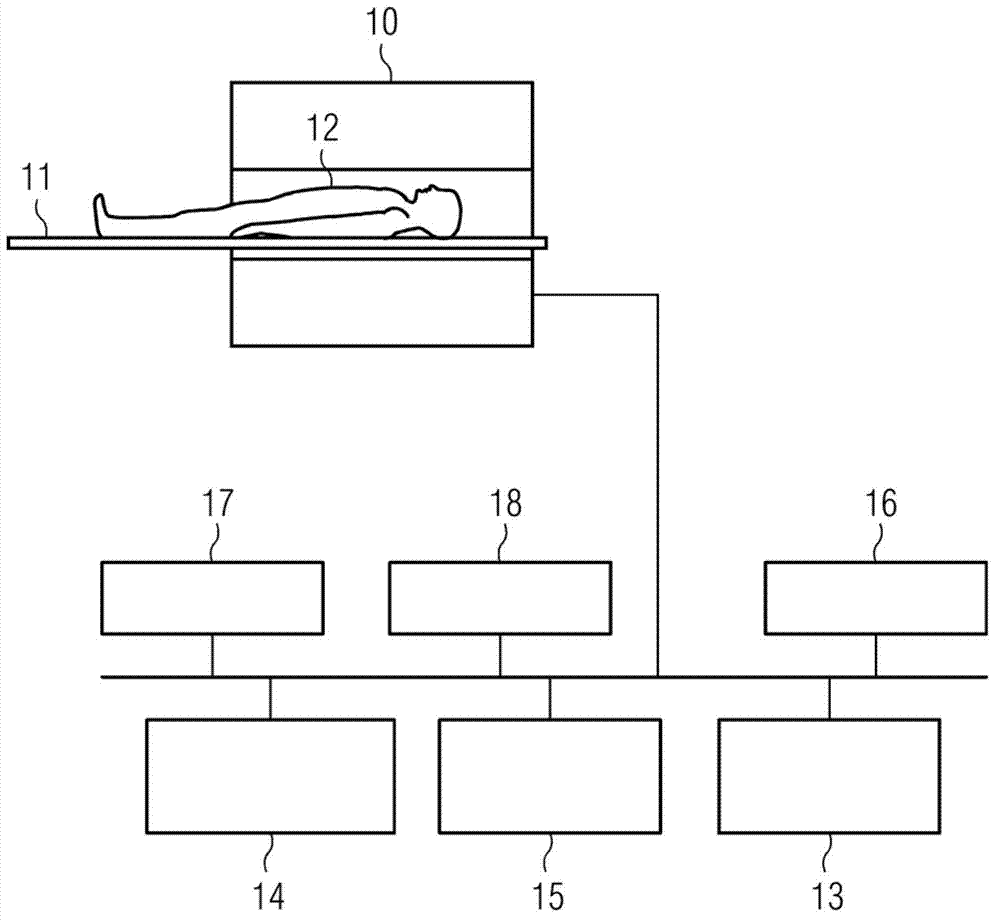
[0034] exist figure 1 , schematically shows an MR system with which MR angiography images with good spatial resolution can be generated in all three spatial directions within an acceptable measurement time. The MR system has a magnet 10 for generating a polarization field B0. A person to be examined 12 arranged on a couch 11 is moved into the magnet 10 , wherein the magnetization generated in the person being examined can be reversed out of the equilibrium position by radiating high-frequency pulses. The relaxation processes that occur after the incident radio-frequency pulse can be detected with coils not shown. For the spatial encoding of the detected signals, magnetic field gradients are also applied via gradient coils (not shown) in order to achieve a spatial dependence of the detected signals. The general method of how MR images can be generated by the sequence of radiating high-frequency pulse sequences and switching on magnetic field gradients is known to those skille...


PUM


Abstract

In a method and apparatus for the creation of an MR image of a vascular structure of an examination region, the spins in the examination region are saturated by the irradiation of at least one RF saturation signal, which delivers a lower signal intensity as spins in a subsequent MR signal recording for the creation of the MR angiographic image, which flow through at least one blood vessel into the examination region, and are not saturated by the RF saturation pulse. Raw data space of the MR angiographic image is read out with a non-Cartesian trajectory in the MR signal acquisition for the creation of the MR angiographic image.

Description

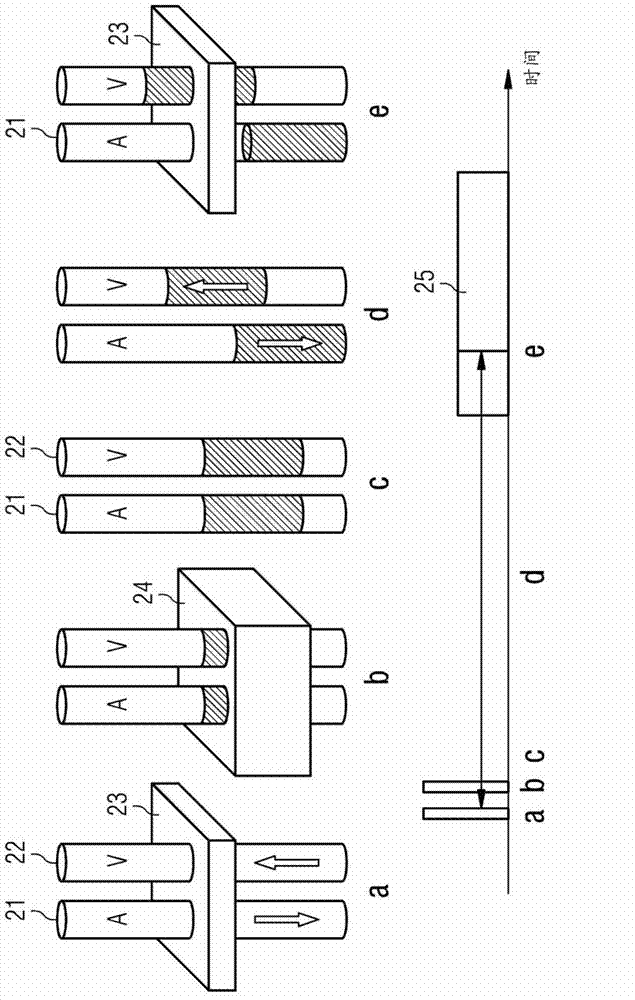
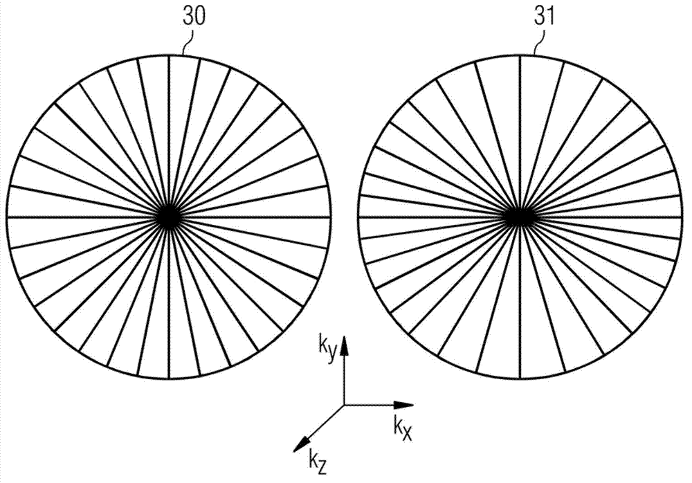
technical field [0001] The invention relates to a method for generating an MR image of a vessel structure of an examination region and an MR system for the method. The term “angiographic image” is used generally below: it does not necessarily describe only a single two-dimensional image, but, depending on the context, optionally also an angiographic image dataset of a desired volume. Background technique [0002] In the field of MR angiography, methods that work without contrast agents are gaining increasing importance. Methods in this class attempt to take advantage of the pulsatile nature of arterial blood flow. Ideally, extinction of the arterial signal is obtained by signal acquisition with flow-sensitive sequences at moments of rapid flow velocity (systole). By recording the signal at the moment of the cardiac cycle (diastole) at which the velocity is at a minimum or zero, ideally an artery with a bright signal is obtained. By subtracting the two recorded data, the s...

Claims


Application Information

Patent Timeline
Patent Type & Authority Applications(China)
IPC IPC(8): G01R33/563A61B5/055
CPCG01R33/5635G01R33/4824
Inventor P.施米特
Owner SIEMENS HEALTHCARE GMBH
Learn More