Method for extracting microwave detection breast model based on medical magnetic resonance imaging

A nuclear magnetic resonance image and nuclear magnetic resonance technology, applied in the field of biomedical detection, can solve the problems of human radiation damage, low imaging contrast, and high cost

Active Publication Date: 2014-02-05
TIANJIN UNIV
View PDF4 Cites 18 Cited by
  • Summary
  • Abstract
  • Description
  • Claims
  • Application Information

AI Technical Summary

Problems solved by technology

Currently commonly used detection methods for early breast cancer include mammography, ultrasound imaging technology, computed tomography, nuclear magnetic resonance imaging technology, thermal imaging detection, etc., but many methods have certain shortcomings, such as radiation damage to the human body, Low imaging contrast, high cost, etc.

Method used


Image

  • Method for extracting microwave detection breast model based on medical magnetic resonance imaging
  • Method for extracting microwave detection breast model based on medical magnetic resonance imaging
  • Method for extracting microwave detection breast model based on medical magnetic resonance imaging

Examples


Embodiment Construction

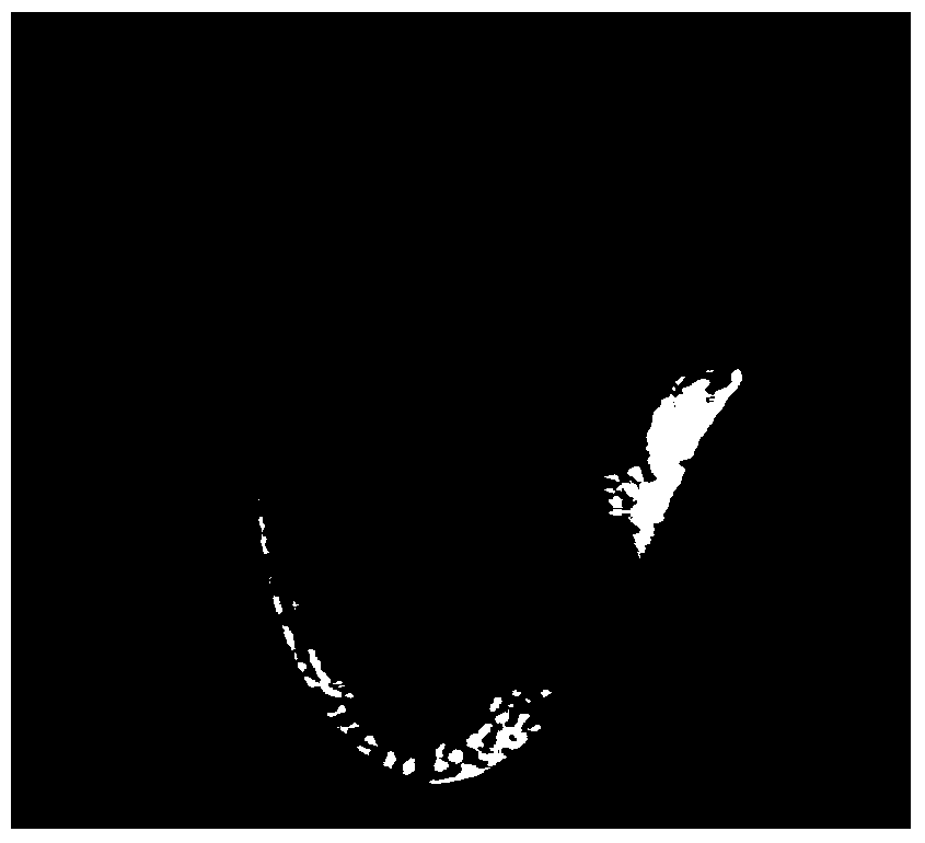
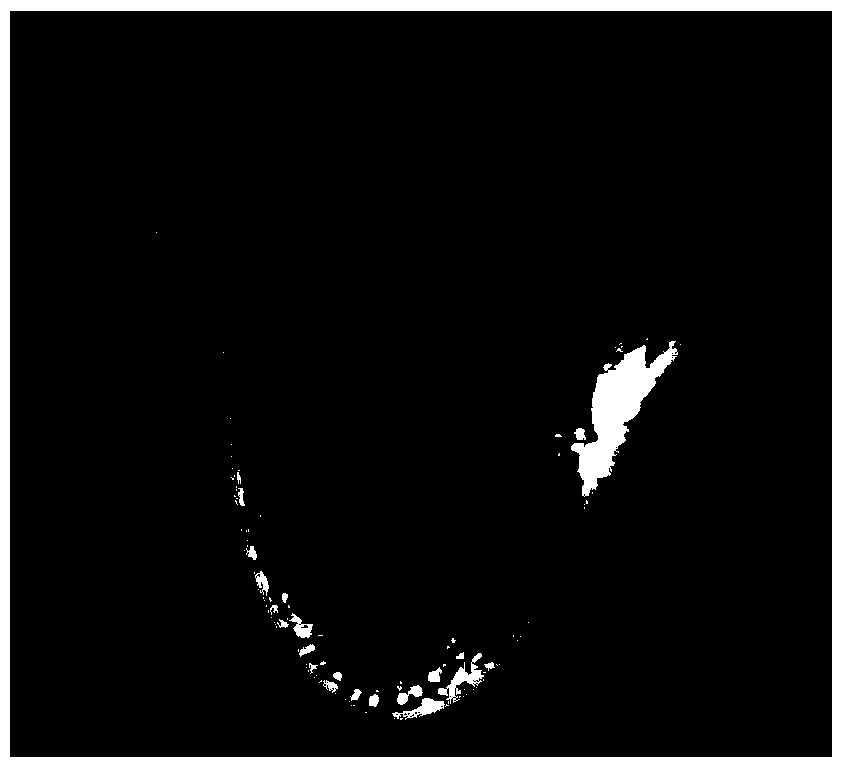
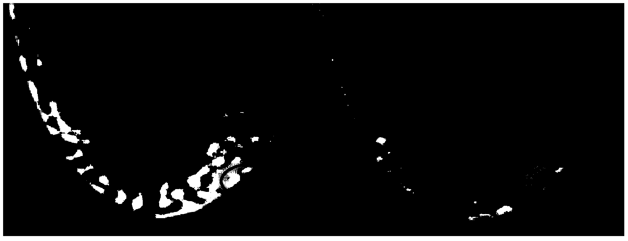
[0032] The present invention mainly performs processing based on the grayscale of the MRI image and its image morphological features. Realize the function of extracting the specific shape and position of each tissue in the breast from the MRI image, and finally convert it into the corresponding standard storage format according to the simulation requirements. In this way, a more realistic model can be provided for subsequent electromagnetic simulations, and a good simulation comparison and reference can also be provided for clinical measurements. It is mainly divided into the following four steps:

[0033] 1. Boundary Enhancement

[0034] In traditional MRI images, due to the limitations of noise, low image resolution, etc., it is not very obvious to distinguish the boundaries of various tissues in the breast. Although the gray value of each tissue in the MRI image is different, because the boundaries are not obvious, it is impossible to distinguish the boundaries of various ...


PUM


Abstract

The invention belongs to the technical field of biomedical detection, and relates to a method for extracting a microwave detection breast model based on medical magnetic resonance imaging. The method includes the steps: acquiring a T1 weighted magnetic resonance image of a breast portion; enhancing a boundary; performing gray discrete processing; calculating the water content of a tissue corresponding to each pixel for a thoracic gland portion in the magnetic resonance image by the aid of an image gray value; comparing the water content of the tissue corresponding to each pixel with typical water content data of each tissue of a normal human body, determining tissue information of each pixel and defining the pixel as an islet if the tissue information of the pixel cannot be determined; giving the same gray value to the pixels with the determined tissue information and belonging to the same tissue, and normalizing the gray of the magnetic resonance image according to the tissue information; corroding small discrete islets; building the breast model. A breast structure can be really simulated by the method.

Description

Technical field [0001] The invention belongs to the technical field of biomedical detection, and relates to a method for extracting a microwave detection model. Background technique [0002] Breast cancer is the malignant tumor disease with the highest incidence rate in women, and its mortality rate ranks first in the mortality rate of malignant tumors in women. The diagnosis of early breast tumors is undoubtedly of decisive significance for improving the treatment rate of breast disease and the long-term survival rate of patients. Currently commonly used detection methods for early breast cancer include mammography, ultrasound imaging technology, computed tomography, nuclear magnetic resonance imaging technology, thermal imaging detection, etc., but many methods have certain shortcomings, such as radiation damage to the human body, The imaging contrast is low and the cost is high. The principle of ultra-broadband electromagnetic wave detection of breast cancer is that dif...

Claims


Application Information

Patent Timeline
Patent Type & Authority Applications(China)
IPC IPC(8): A61B5/055G01R33/56
Inventor 肖夏宋航王宗杰
Owner TIANJIN UNIV
Why Eureka
Try Eureka