Vascular imaging method and device

A technology of blood vessels and blood vessel paths, applied in the field of medical images, can solve the problems that subtraction images cannot accurately and completely display blood vessel areas, motion displacement, subtraction, etc.

Active Publication Date: 2014-06-25
BEIJING NEUSOFT MEDICAL EQUIP CO LTD
View PDF9 Cites 15 Cited by
  • Summary
  • Abstract
  • Description
  • Claims
  • Application Information

AI Technical Summary

Problems solved by technology

The implementation of the existing subtraction technology requires two scans of the patient, and in the second scan, the patient needs to be injected with a contrast agent, which results in a longer time interval between the first scan and the second scan
Since it is difficult for the patient to maintain the same body position for a long time, the detected area

Method used


Image

  • Vascular imaging method and device
  • Vascular imaging method and device
  • Vascular imaging method and device

Examples


Embodiment Construction

[0065] In order to make the above objects, features and advantages of the present invention more comprehensible, the embodiments of the present invention will be described in detail below in conjunction with the accompanying drawings.

[0066] It should be noted that the method of the present invention can be used in systems for scanning and displaying blood vessels, such as: CT (Computed Tomography) electronic computer X-ray scanning system, PET-CT (Positron Emission Tomography) positron emission tomography scanning system, MRI ( Magnetic Resonance Imaging) magnetic resonance imaging system and other systems.

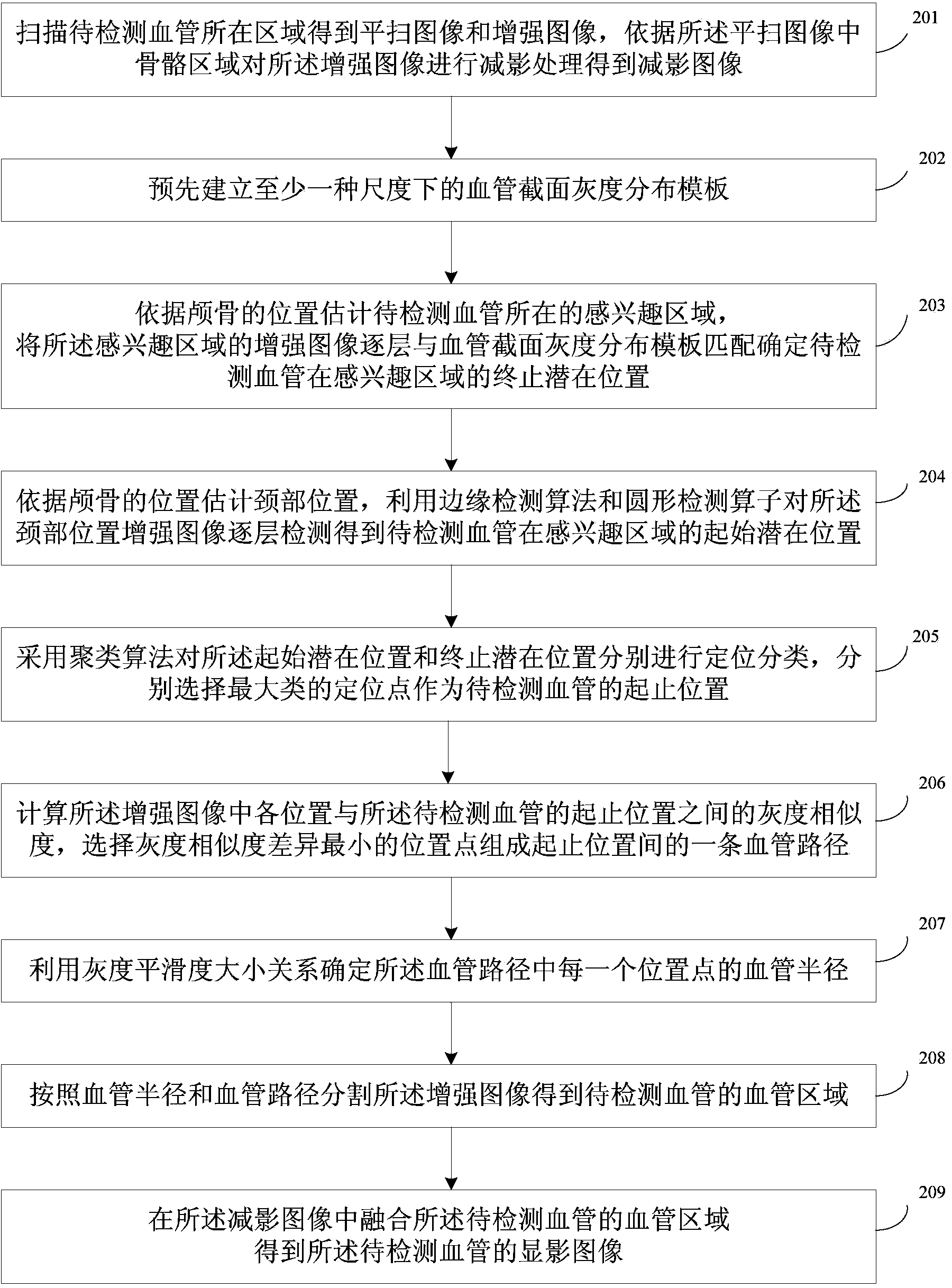
[0067] see figure 1 , shows the flow chart of Embodiment 1 of the blood vessel imaging method according to the embodiment of the present invention, and the method includes: step 101-step 103.

[0068] Step 101, scan the region where the blood vessel to be detected is located to obtain a plain scan image and an enhanced image, and perform subtraction processing on the ...


PUM


Abstract

Embodiments of the invention disclose a vascular imaging method and device. The method includes: scanning an area of blood vessels to be detected to obtain a plain scan image and an enhanced image, and subtracting the enhanced image according to a skeletal area in the plain scan image to obtain a subtracted image; detecting a vascular area of the blood vessels to be detected, in the enhanced image; fusing the vascular area of the blood vessels to be detected, in the subtracted image to obtain an image of the blood vessels to be detected. Therefore, by the use of the vascular imaging method and device, the blood vessels passing skeleton can be retained in the subtracted image.

Description

technical field [0001] The invention relates to the technical field of medical images, in particular to a blood vessel developing method and device. Background technique [0002] Vascular disease is one of the diseases that seriously affect human health. Therefore, the requirements for vascular imaging technology are getting higher and higher. Due to the complex shape and structure of blood vessels in the human body, some blood vessels are blocked by bones, and it is difficult to display them intuitively in the 3D volume rendering results. The overall shape and structure of blood vessels. For example: head and neck blood vessels start from the heart and enter the brain through the bone area at the bottom of the skull, carotid arteries pass through the skull, vertebral arteries pass through the six vertebrae and enter the interior of the skull, etc. [0003] At present, the subtraction technique is usually used to subtract the bone area in the image so as to preserve all the...

Claims


Application Information

Patent Timeline
IPC IPC(8): A61B6/03A61B5/055
CPCA61B5/055A61B5/489A61B5/7425A61B5/748A61B6/032A61B6/481A61B6/504A61B6/5217A61B6/5241G16H50/30A61B6/037A61B6/466A61B6/469A61B6/501A61B6/5252
Inventor 赵建张博闻
Owner BEIJING NEUSOFT MEDICAL EQUIP CO LTD
Why Eureka
Try Eureka