Cells for clearing amyloid polypeptide and application thereof
An amyloid polypeptide, cell technology, applied in the field of cell biology and pharmacy, can solve problems such as cell dysfunction
- Summary
- Abstract
- Description
- Claims
- Application Information
AI Technical Summary
Problems solved by technology
Method used
Image
Examples
Embodiment 1
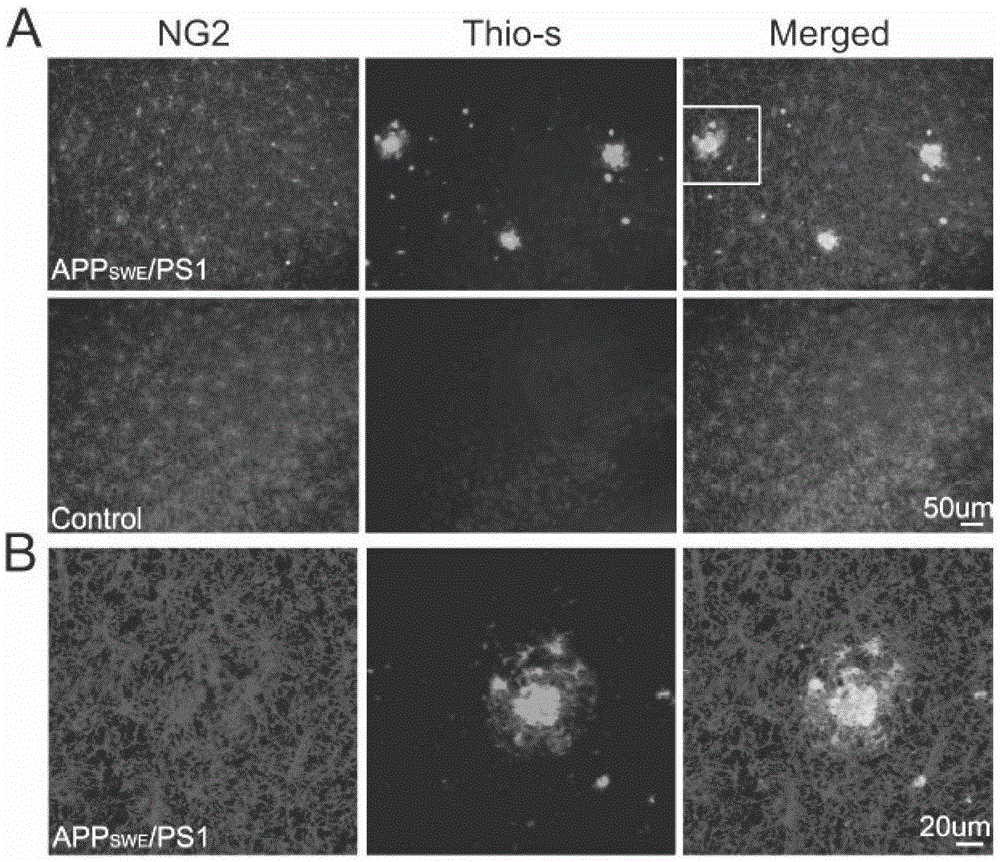
[0199] Example 1, NG2 cells are activated around amyloid plaques
[0200] The brain tissue sections (brain slices) of 14-month-old APPswe / PS1 transgenic mice (JacksonLab, clone number 004462) and control mice without APPswe / PS1 transgene (C57BL / 6J*C3H / HeJ) were obtained, and the NG2 cells of the cerebral cortex were analyzed. Immunohistochemical staining and plaque thioflavin-S staining. NG2-positive cells were uniformly distributed in the cerebral cortex of control mice, but gathered around amyloid plaques in APPswe / PS1 transgenic mice, as shown in figure 1 A-B are shown.
Embodiment 2
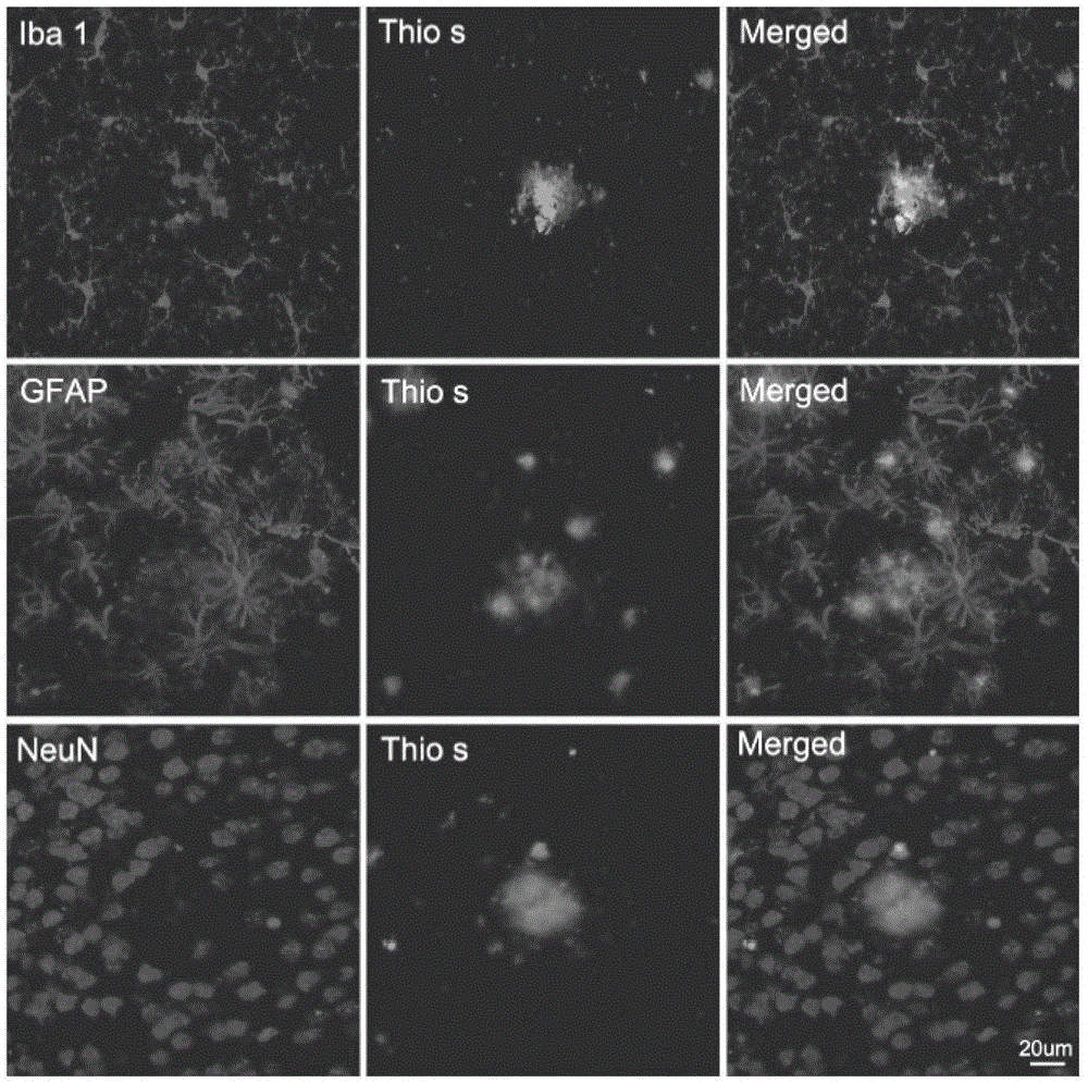
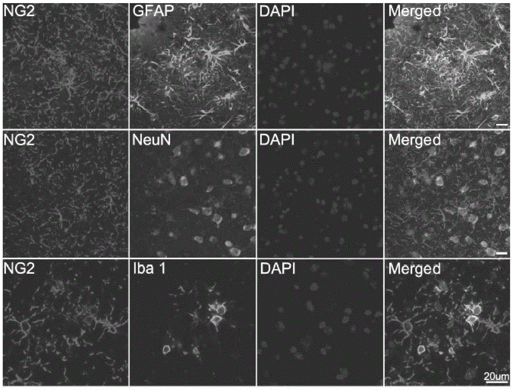
[0201] Example 2, NG2 cells are a new cell type that gathers around amyloid plaques
[0202] Immunohistochemical staining of the cerebral cortex of APPswe / PS1 transgenic mice. Such as figure 2 , Thioflavin-S staining for amyloid plaques, Iba1 for microglia, GFAP for astrocytes, and NeuN for neurons. The results showed that Iba1-positive microglia and GFAP-positive astrocytes gathered around the amyloid plaques, while there were no NeuN-positive neurons at the location of the amyloid plaques.
[0203] Immunohistochemical double labeling was performed on the cerebral cortex of APPswe / PS1 transgenic mice. The cerebral cortex was double-labeled with NG2 and GFAP, NeuN or Iba1. The result is as image 3 , showing that NG2-positive cells did not co-localize with GFAP-positive astrocytes, NeuN-positive neurons and Iba1-positive microglia. The results demonstrated that NG2 cells are a new cell type that aggregates around amyloid plaques, where they are activated.
Embodiment 3
[0204] Example 3, primary NG2 cells and oli-neu cell lines can phagocytize Aβ
[0205] The primary NG2 cells and Oli-neu cells were seeded on slides in a 24-well plate, cultured for 18 hours, and then incubated with 2 μM fluorescently-labeled (HiLyteFluor488-labeled) Aβ for 24 hours. Cell immunofluorescence staining with NG2 antibody was performed after the cells were fixed. The result is as Figure 4 A-B, showing that Aβ can be phagocytosed into the somata of primary NG2 cells and Oli-neu cells.
PUM
 Abstract
Description
Claims
Application Information
 - R&D
- Intellectual Property
- Life Sciences
- Materials
- Tech Scout
- Unparalleled Data Quality
- Higher Quality Content
- 60% Fewer Hallucinations
Browse by: Latest US Patents, China's latest patents, Technical Efficacy Thesaurus, Application Domain, Technology Topic, Popular Technical Reports.
© 2025 PatSnap. All rights reserved.Legal|Privacy policy|Modern Slavery Act Transparency Statement|Sitemap|About US| Contact US: help@patsnap.com


