
X-ray medical image objective reconstruction based on independent component analysis

An independent component analysis and medical image technology, which is applied in the field of biomedical engineering, can solve problems such as low contrast, difficult to distinguish, and large noise, and achieve the effects of improving image signal-to-noise ratio, accurate detection, and improving material identification accuracy

Inactive Publication Date: 2014-10-08
NANJING UNIV OF POSTS & TELECOMM
View PDF1 Cites 6 Cited by
  • Summary
  • Abstract
  • Description
  • Claims
  • Application Information

AI Technical Summary

Problems solved by technology

Based on the different attenuation of X-rays by different tissue components, multiple fluoroscopic images with different optical density distributions can be obtained, but there are problems such as large noise and low contrast, and human tissue images are superimposed on each other and it is difficult to distinguish them.

Method used


Image

  • X-ray medical image objective reconstruction based on independent component analysis
  • X-ray medical image objective reconstruction based on independent component analysis
  • X-ray medical image objective reconstruction based on independent component analysis

Examples


Embodiment Construction

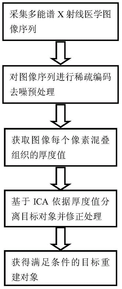
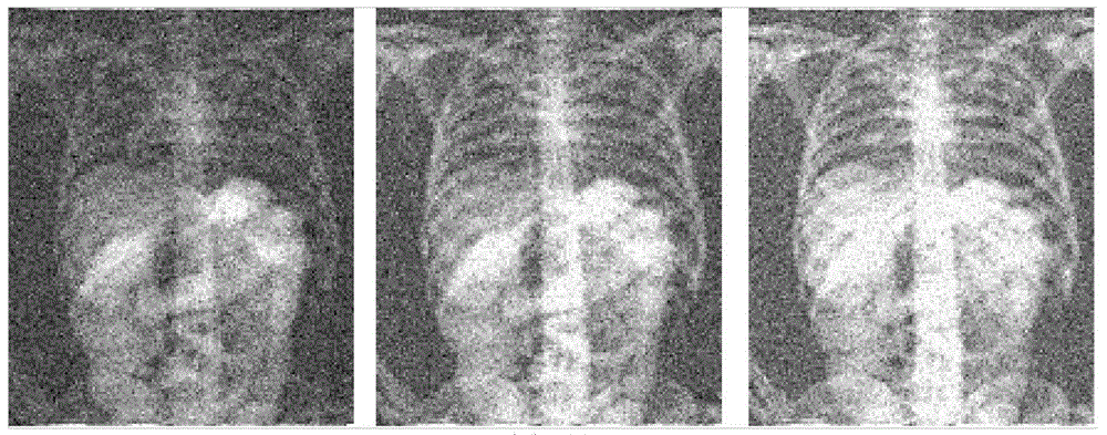
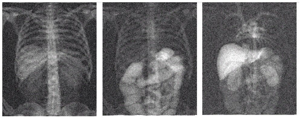
[0022] The X-ray medical image target reconstruction method based on independent component analysis, in order to reduce the difficulty of reconstruction, the image is directly denoised and preprocessed to meet the precondition of independent component analysis separation; then, each pixel is separated according to the difference in the proportion and thickness of the main components of the photographed organs The aliasing thickness value; based on the optimization and improvement of Fast Independent Component Analysis (FastICA) with better convergence speed and separation effect, it meets the medical image reconstruction conditions; finally, the separated target image is corrected according to the subjective evaluation.

[0023] When the X-ray beam passes through the human body, the human body absorbs the X-ray to different degrees, and the energy of the X-ray beam passing through different tissues to the detector is also different. Assume that the initial energy of X-rays in t...


PUM


Abstract

The invention discloses an X-ray medical image objective reconstruction method based on independent component analysis. In order to solve the problems that a traditional medical X-ray image is high in noise and poor in layering, and organs and tissue overlap, image denoising and objective extraction are carried out by combining a multi-energy-spectrum X-ray imaging technology and an independent component analysis algorithm. The method comprises the steps that first, denoising preprocessing is carried out on a medical image so that the preconditions for objective separation through independent component analysis can be met; second, the thickness value of an aliasing organ in each pixel is obtained according to an X-ray attenuation energy matrix of organ and tissue; third, the independent component analysis algorithm is used for adjusting the frequency of convergence and the size of signal scales according to the thickness value of the aliasing organ, a convergence matrix is obtained, the image of each organ is separated, contrast ratio correction is carried out according to an subjective vision standard, an area of interest and marginal information stand out, and the visual and clear image applicable to medical analysis are obtained.

Description

technical field [0001] The invention belongs to the research field of image signal separation and digital image processing in the field of biomedical engineering, and specifically refers to the target extraction and reconstruction of multi-energy spectral X-ray medical images using an improved independent component analysis algorithm. Background technique [0002] Since Roentgen discovered X-rays in 1895, X-rays have been widely used in medical imaging. The purpose of X-ray imaging is to enable doctors to clearly observe a diseased tissue in the patient's body, so the quality of medical images directly affects the accuracy of medical diagnosis. In the 1970s, Robert E. Alvarez and Albert Macovski combined dual-energy imaging with X-ray energy spectrum information to initially realize the identification of material components, which was initially used for the separation of bones and soft tissues and the diagnosis of lesions in medical radiographic images. However, the discrim...

Claims


Application Information

Patent Timeline
Patent Type & Authority Applications(China)
IPC IPC(8): G06T11/00
Inventor 喻春雨李艳缪亚健费彬
Owner NANJING UNIV OF POSTS & TELECOMM
Learn More