Neuron-like cell sourced from humanized adipose-derived stem cells, preparation method and application thereof
A technology of neuron-like cells and adipose-derived stem cells, which is applied in the field of stem cells and biomedicine, and can solve problems such as little consideration of clinical transformation applications, toxicity to the human body, and low maturity of neuron-like cells.
- Summary
- Abstract
- Description
- Claims
- Application Information
AI Technical Summary
Problems solved by technology
Method used
Image
Examples
Embodiment 1
[0099] Example 1. Characterization of hADSCs by cell morphology and CD surface markers
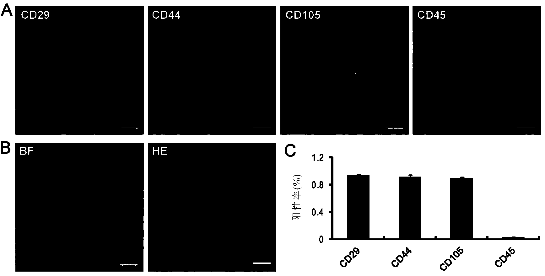
[0100] The tissue cells obtained from liposuction were diluted with 20% fetal bovine serum (JRH Bioscience, USA) basal medium DMEM / F-12 (Invitrogen, Japan) and seeded in T-75flasks (Falcon, Becton Dickinson, Japan). ), so that its concentration is 6×10 7 cells / cm 2 . The medium was changed every two weeks. After 15 days of culture, hADSCs adhered to the wall, showed a typical spindle shape, and expanded in a vortex manner. Hematoxylin and eosin (HE) staining showed that hADSCs were mononuclear cells, such as figure 1 Shown in B. Identify hADSCs by immunohistochemical staining for CD surface markers, such as figure 1 a. Quantification of the positive rate of CD surface markers as figure 1 c.
[0101] These results indicated that more than 85% of hADSCs highly expressed the mesenchymal stem cell markers CD29, CD44, and CD105, whereas less than 5% of hADSCs expressed the hematopoieti...
Embodiment 2
[0102] Example 2. Stem cell transcription factors and differentiation to three lineages indicate pluripotency of hADSCs
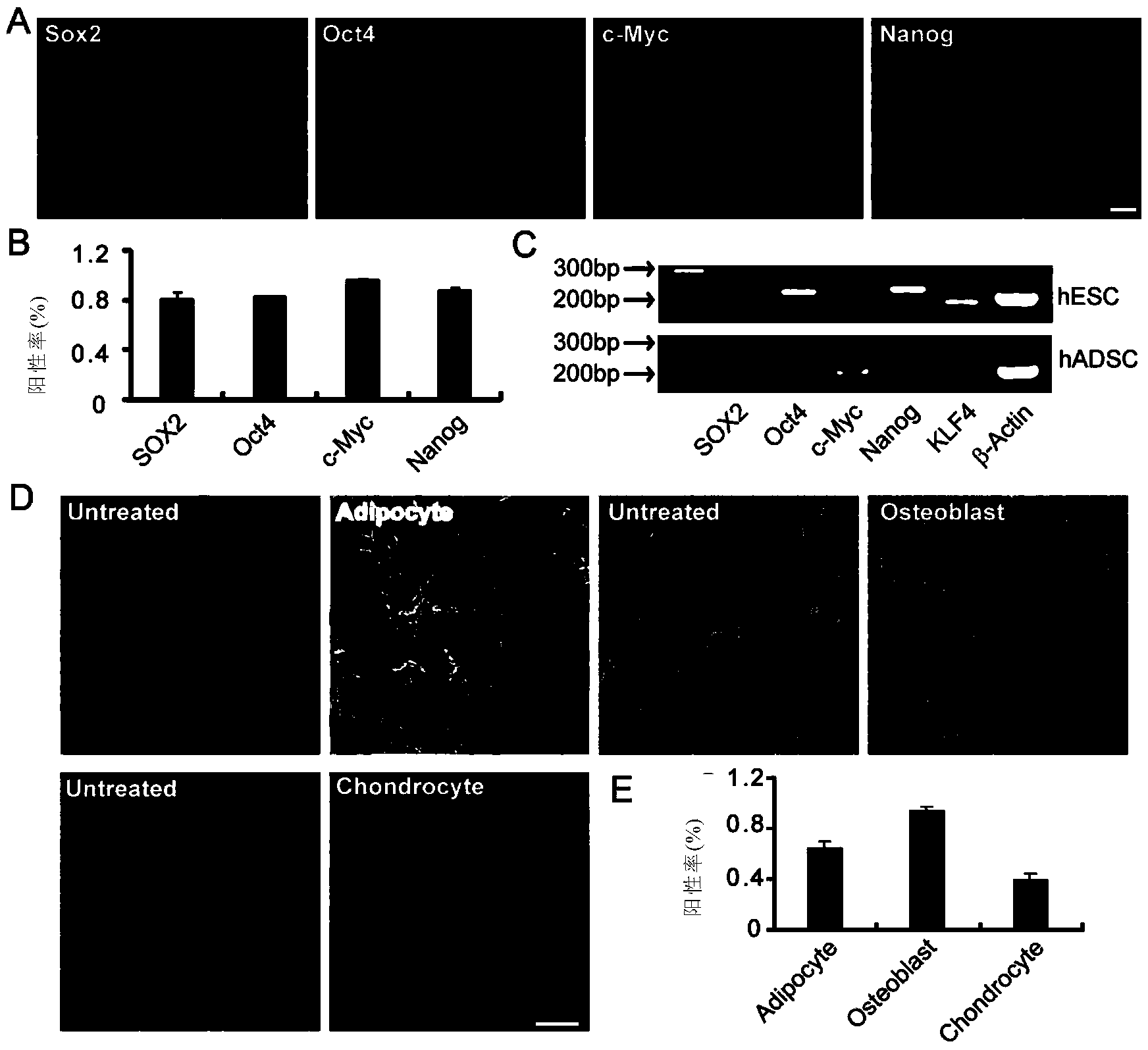
[0103] Classic stem cell transcriptional markers such as Sox2, Oct4, c-Myc, Nanog, and KLF4 have been widely used to characterize induced pluripotent stem cells (iPSCs). To characterize the stemness of hADSCs, this study applied immunohistochemical staining and designed primers for transcription factor markers and semi-quantitative RT-PCR to detect the expression of these factors, compared with ESCs. The results showed that: with immunohistochemical staining, hADSC expressed Sox2, Oct4, c-Myc and Nanog at the protein level ( figure 2 A). Human embryonic stem cell (hESC) line H9 was used as a positive control, and Sox2, Oct4, c-Myc, Nanog and KLF4 were expressed by semi-quantitative RT-PCR, and Oct4, Nanog and KLF4 were significantly highly expressed in hESC. The number of positively expressed stem cell transcriptional markers indicated that more than 80%...
Embodiment 3
[0104] Example 3. Differentiation of hADSCs to neurons in plastic culture dishes into hADSC-NLCs
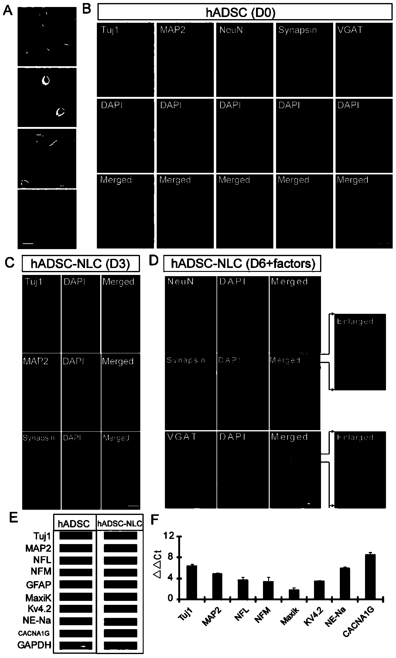
[0105] The inventors induced hADSCs to differentiate into neurons into hADSC-NLCs in plastic culture dishes. The steps of neuron induction included: first incubating hADSCs with DKK1 for 24 hours, and then using Safford, K.M, etc. (Biochem Biophys Res Commun294, 371, 2002) described in the mixed medium culture for 3 days. During induction, hADSC morphology changes, the cytoplasm retracts toward the nucleus, and the cell body becomes spherical, as in image 3 A and B are shown.
[0106] In order to confirm that the expression of neuronal markers is really due to the induction of the mixed induction solution, hADSCs were first immunohistochemically stained with Tuj1, MAP2, NeuN, Synapsin1 / 2 and vGAT without any treatment. hADSCs basally express Tuj1 and MAP2 but not NeuN, Synapsin1 / 2 and vGAT, as image 3 C shown. Basal expression of some neuronal markers in hADSCs, such as Tuj...
PUM
 Abstract
Description
Claims
Application Information
 - R&D
- Intellectual Property
- Life Sciences
- Materials
- Tech Scout
- Unparalleled Data Quality
- Higher Quality Content
- 60% Fewer Hallucinations
Browse by: Latest US Patents, China's latest patents, Technical Efficacy Thesaurus, Application Domain, Technology Topic, Popular Technical Reports.
© 2025 PatSnap. All rights reserved.Legal|Privacy policy|Modern Slavery Act Transparency Statement|Sitemap|About US| Contact US: help@patsnap.com


