Handheld scanning device

A scanning device and a handheld technology, applied in medical science, image data processing, 3D image processing, etc., can solve problems such as incorrect diagnosis, difficult reconstruction, and failure to observe the image plane, to achieve enhanced exposure, ultrasound Optimum image quality

Inactive Publication Date: 2015-01-21
安华亿能医疗影像科技(北京)有限公司
View PDF16 Cites 14 Cited by
  • Summary
  • Abstract
  • Description
  • Claims
  • Application Information

AI Technical Summary

Problems solved by technology

[0003] 1. Conventional ultrasound images are two-dimensional, and the diagnostician must mentally transform multiple two-dimensional images to form a complex three-dimensional structure, which leads to variability and sometimes incorrect diagnoses;
[0004] 2. It is very difficult to localize the 2D ultrasound image plane of the organ and reconstruct it afterwards
This makes the approach error-prone and limited in its ability to quantify disease progression and decline, especially when tracking small changes;
[0005] 3. The patient's body structure often limits the angle of the image, resulting in the inability to observe the most ideal image plane for disease assessment;
Whereas 2D ultrasound is variable and sometimes inaccurate, 3D ultrasound ensures accurate and less variable estimation of anatomy

Method used


Image

  • Handheld scanning device
  • Handheld scanning device
  • Handheld scanning device

Examples


Embodiment approach

[0100] a) Magnetic tracker: One or more FM transmitters are used to generate a magnetic field that varies with space, and one or more FM receivers containing three-phase quadrature coils are used to sense the strength of the magnetic field. The position and orientation information of the transducer can be calculated by tracking the magnetic field strength of the three phases generated by the FM transmitter every time a two-dimensional slice image of the carotid artery is obtained.

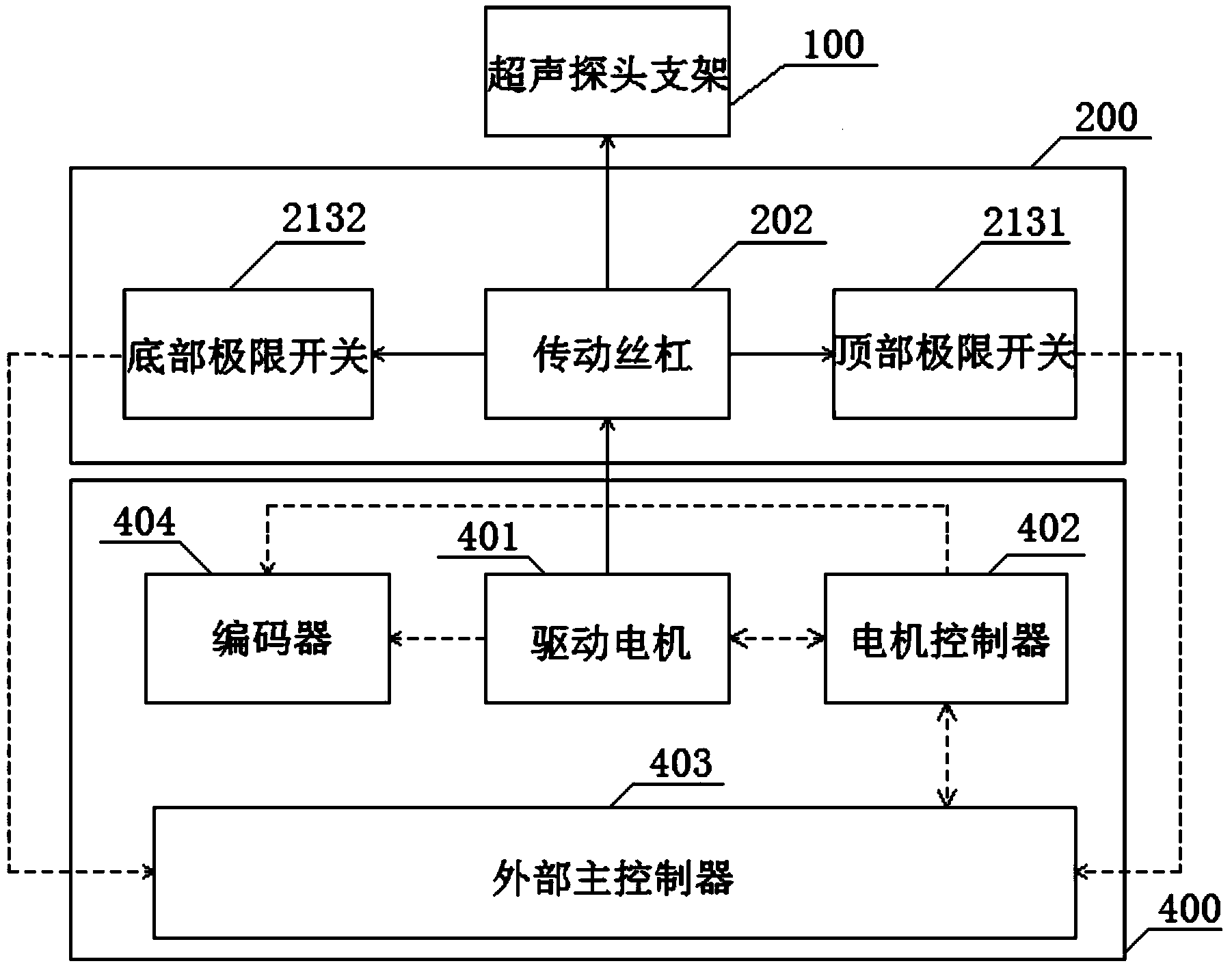
[0101] b) Mechanical Tracker: The drive mechanism in this approach is preferentially controlled by a drive motor or mechanical system and operates at a constant and predictable rate. It then also employs a spring-balanced mechanism or automatic mechanical clamping mechanism. Depending on the imaging system, the mechanical tracker is driven in the following configurations: 1) linear - where images are acquired parallel to each other with equal or dynamic spacing; 2) oblique - in a fan-like configura...


PUM


Abstract

The invention discloses a handheld scanning device. The handheld scanning device comprises an auxiliary handheld scanning device, an ultrasonic probe and a drive control device. The auxiliary handheld scanning device comprises an H-shaped hollow shell, a scanning transmission mechanism fixed to the hollow shell, and an ultrasonic probe support in transmission connection with the scanning transmission mechanism. The ultrasonic probe is fixed to the ultrasonic probe support. The drive control device comprises a drive motor, a motor control box and an external master controller. The external master controller carries out driving control on the drive motor through the motor control box. The drive motor drives the scanning transmission mechanism to drive the ultrasonic probe support to move in a linear mode so that the ultrasonic probe can continuously and stably collect two-dimensional carotid artery section images at different positions. The handheld scanning device can continuously, accurately and stably collect the two-dimensional ultrasonic images.

Description

technical field [0001] The invention relates to the technical field of image acquisition and processing, in particular to a hand-held scanning device. Background technique [0002] Medical ultrasound imaging technology is today's mainstream method for easy and non-invasive assessment of the detailed internal structure of tissues and organs for assessment, diagnosis, designation of treatment options, and determination of curative effect. However, conventional 2D ultrasound image scanning has several drawbacks: [0003] 1. Conventional ultrasound images are two-dimensional, and the diagnostician must mentally transform multiple two-dimensional images to form a complex three-dimensional structure, which leads to variability and sometimes incorrect diagnoses; [0004] 2. It is very difficult to localize the 2D ultrasound image plane of the organ and reconstruct it afterwards. This makes the approach error-prone and limited in its ability to quantify disease progression and dec...

Claims


Application Information

Patent Timeline
Patent Type & Authority Applications(China)
IPC IPC(8): A61B8/00G06T15/00
CPCA61B8/00
Inventor 汪洋魏桑迪
Owner 安华亿能医疗影像科技(北京)有限公司
Why Eureka
Try Eureka