Fever to-be-checked computer aided diagnosis method based on PET/CT images

A computer-aided, CT imaging technology, applied in computing, image enhancement, image analysis, etc., can solve the problem that it is difficult to deal with uneven lesion tissue and diffuse lesion tissue, the whole body bones cannot achieve good segmentation results, and it is difficult to accurately segment The whole body bone marrow tissue and other problems, to achieve the effect of improving algorithm robustness, shortening patient diagnosis time, and wide applicability

Inactive Publication Date: 2015-03-25
BEIJING INSTITUTE OF TECHNOLOGYGY
View PDF3 Cites 24 Cited by
  • Summary
  • Abstract
  • Description
  • Claims
  • Application Information

AI Technical Summary

Problems solved by technology

[0007] 1. The current bone segmentation methods are all designed for extracting the outline of the bone. The bone marrow has a lower CT value than the bone cortex. It is difficult for the existing methods to accurately segment the bone marrow tissue of the whole body.
[0008] 2. The existing bone segmentation methods for CT images are all designed for a certain bone. The adaptability and robustness of the algorithm are relatively poor, and good segmentation results cannot be achieved when segmenting the whole body bones.
[0009] 3. Due to differences in imaging principles, the resolution of PET images is much lower than that of CT images
[0010] 4. The existing indicators for evaluating PET images are mainly the average value of the region of interest, which is difficult to deal with heterogeneous lesion tissue and diffuse lesion tissue
However, the existing analysis methods for PET / CT fusion images are very limited, mainly relying on doctors to observe and analyze limited regions of interest, so they are highly subjective
The clinical diagnosis process often requires doctors to have strong professional knowledge and a lot of operating experience

Method used


Image

  • Fever to-be-checked computer aided diagnosis method based on PET/CT images
  • Fever to-be-checked computer aided diagnosis method based on PET/CT images
  • Fever to-be-checked computer aided diagnosis method based on PET/CT images

Examples


Embodiment Construction

[0034] The present invention will be further described below in conjunction with the accompanying drawings.

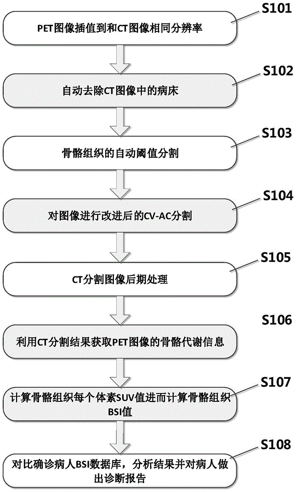
[0035] attached figure 1 To reconstruct the flowchart, the diagnosis consists of the following steps:

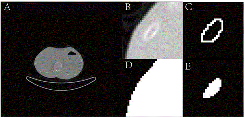
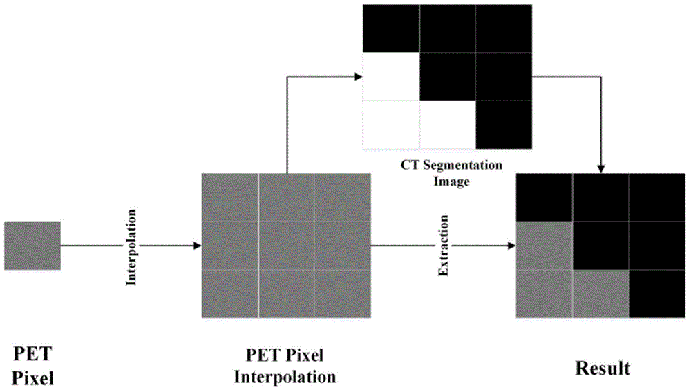
[0036]Step S101, the resolution of the original CT tomographic image is A×A, and the resolution of the original PET tomographic image is B×B. Due to the different imaging principles, the resolution of CT images is much higher than that of PET images (A>B) . Before extracting PET voxels, PET images and CT images need to be converted to the same resolution. Because reducing the CT image resolution to PET image resolution will seriously affect the accuracy of CT bone segmentation, we choose to interpolate the PET image resolution from B×B to A×A. In order to keep all the information of the PET image as much as possible, we use the nearest neighbor interpolation method for interpolation. This method interpolates a PET pixel into n×n pixels with the same gray value. I...


PUM


Abstract

The invention provides a fever to-be-checked computer aided diagnosis method based on PET/CT images. Full-automatic analysis of the PET/CT bone images is achieved, and a doctor is assisted in diagnosing fever to-be-checked patients. Firstly, lossless interpolation of the PET images is conducted; secondly, hospital beds in the CT images are automatically removed; thirdly, full-automatic bone segmentation of the CT images is conducted; fourthly, bone segmentation is conducted on the whole-body CT images through an optimal active contour model of a CV active contour region; fifthly, after-treatment of the CT segmented images is conducted; sixthly, bone tissue information of the PET images is acquired; seventhly, bone tissue SUVs and bone tissue BSI values are acquired through calculation, wherein the SUVs of bones, marrow and bone cortices are acquired through calculation respectively, and the BSI values of the bones, the marrow and the bone cortices of the patients are acquired through calculation via the SUVs; eighthly, diagnosis is conducted based on the BSI values, wherein the SUVs and the BSI values are compared with SUVs and BSI values in an existing confirmed patient database, and a diagnosis report is made.

Description

technical field [0001] The invention relates to a computer-aided diagnosis method for fever of unknown origin (FUO) based on PET / CT images, which is suitable for the diagnosis and treatment of clinical fever of unknown origin. Background technique [0002] Fever of Unknown Origin (FUO) refers to a group of diseases in which fever lasts for more than 3 weeks, body temperature exceeds 38.3°C several times, and cannot be diagnosed after at least 1 week of in-depth and detailed examination. Despite many new advances in diagnostic techniques, undiagnosed fever remains a major medical problem, with as many as 51% of cases undiagnosed. Among diagnosed patients, there were three main categories, namely infectious disease, malignancy, and noninfectious inflammation. Fluoro-deoxyglucose (Fluoro-18-fluoro-2-deoxy-D-glucose, FDG) is a fluorinated derivative of 2-deoxyglucose, FDG not only accumulates in malignant tissues, but also in infectious diseases and inflammatory tissues Gather...

Claims


Application Information

Patent Timeline
Patent Type & Authority Applications(China)
IPC IPC(8): G06T7/00A61B6/03
CPCA61B6/03G06T7/0012G06T7/11G06T2207/10081G06T2207/10104G06T2207/20116G06T2207/30012
Inventor 杨健刘新新王涌天艾丹妮刘越
Owner BEIJING INSTITUTE OF TECHNOLOGYGY
Why Eureka
Try Eureka