Method and apparatus for increasing field of view in cone-beam computerized tomography acquisition

一种层析成像、计算机的技术,应用在用于放射诊断的仪器、计算、回波层析等方向,能够解决维持FOV尺寸、部件昂贵、经济缺点等问题,达到减少计算时间、缩短获取时间的效果

Active Publication Date: 2015-05-06
CEFLA SOC COOP
View PDF11 Cites 8 Cited by
  • Summary
  • Abstract
  • Description
  • Claims
  • Application Information

AI Technical Summary

Problems solved by technology

In general, each of these solutions involves significant disadvantages: for example, economic disadvantages, because the components are more expensive; or ergonomic disadvantages, related to the need to increase the overall size of the device
[0013] In relation to the possible extension of the imaging FOV according to the solution of US 8363780, although allowing volumetric images to be obtained from larger FOVs, a limitation of FOV size is maintained, which depends on the size of the active area of ​​the sensor and the possible maximum offset
Thus US 8363780 does not propose a solution for further extending the FOV such that it covers the desired ROI in case the extended FOV obtained by the offset of the sensor is still insufficient to cover the entire ROI

Method used


Image

  • Method and apparatus for increasing field of view in cone-beam computerized tomography acquisition
  • Method and apparatus for increasing field of view in cone-beam computerized tomography acquisition
  • Method and apparatus for increasing field of view in cone-beam computerized tomography acquisition

Examples


Embodiment Construction

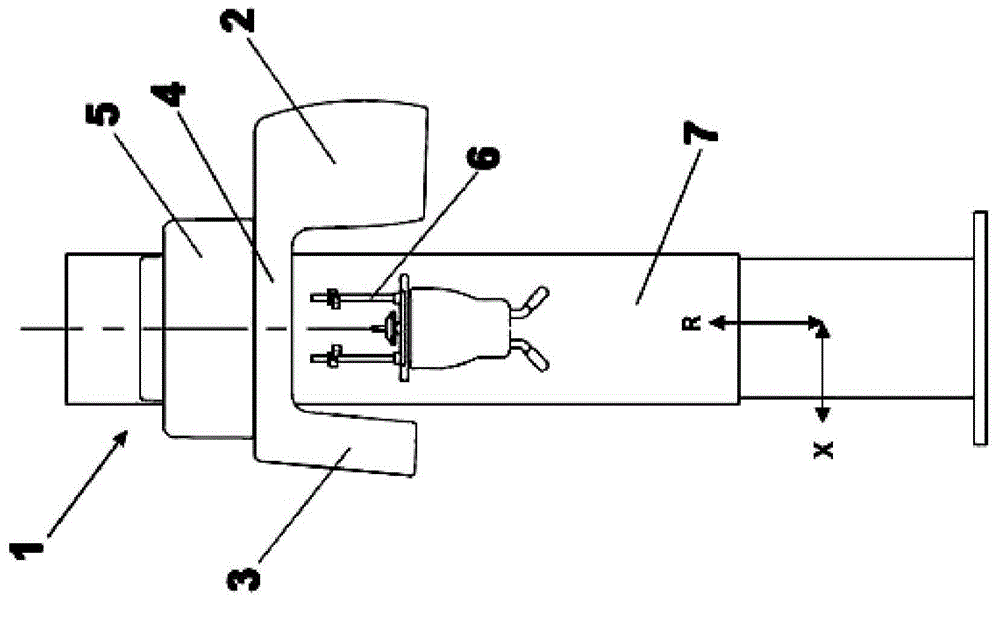
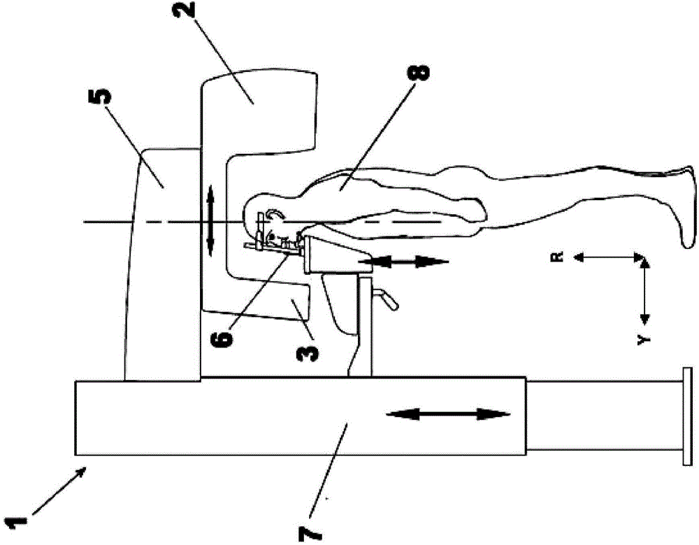
[0061] figure 1 A typical vertical apparatus 1 of known technology is shown, comprising: an X-ray source 2, which projects a cone-beam of X-rays through a patient 8 (unless it is subsequently collimated); a two-dimensional X-ray detector 3, positioned to measure the intensity of radiation after passing through the object; a C-arm on which the X-ray source 2 and detector 3 are fixed; a mechanical system 5 that allows the support to rotate and translate around the patient 8 to move from different position to acquire radiological images; an electronic system (not shown) capable of controlling and synchronizing the operation of the various components of the device; a computer or similar (not shown) capable of allowing the device to be controlled by its user. As mentioned above, the block X-ray sources - the X-ray detectors - and the swivel arm connecting them together are collectively referred to as the beam group 4 . The beam group 44 is provided with three axes of motion: X, Y...


PUM


Abstract

Method and apparatus for increasing the Field Of View dimensions in a cone-beam computerized tomography acquisition, comprising the following steps: A. Selecting an acquisition protocol; B. Positioning a patient; C. Performing a first subacquisition step of a first anatomic area of at least two partially overlapping anatomic areas; D. Repositioning the apparatus for the next subacquisition step; E. Performing the at least one next subacquisition step of the at least further anatomic area; F. Generating the volumetric images relating to the subvolumes; G. Reconstructing a single global volumetric image in which the reconstruction occurs through techniques of image registration (stitching) and blending.

Description

technical field [0001] The present invention relates to a method and a device for cone beam computed tomography (CBCT) based on the acquisition and reconstruction of multiple volumes related to partially overlapping different anatomic regions, and Based on the subsequent stitching of said volumes, the maximum field of view (FOV) can be increased by a compound scanning protocol to obtain as a final result a single final volume with a size larger than the geometry of the acquisition system allows. Background technique [0002] CBCT devices are known in the art and allow obtaining layers of an anatomical part by acquiring a sequence of two-dimensional radiographic images during rotation of a system comprising an X-ray source and an X-ray detector around the anatomical part to be acquired analyze the image. [0003] A CBCT apparatus generally comprises: an X-ray source which projects a cone-shaped beam of X-rays through the object to be acquired (unless it is subsequently colli...

Claims


Application Information

Patent Timeline
Patent Type & Authority Applications(China)
IPC IPC(8): A61B6/03
CPCA61B6/5205A61B6/5241A61B6/54G06T3/4038G06T11/008G06T2200/04G06T2200/32A61B6/4085A61B6/51A61B6/5264A61B6/032A61B6/5211
Inventor G·梅内加兹R·莫尔夏尼B·里瓦尔塔C·山德里F·西蒙内蒂
Owner CEFLA SOC COOP
Why Eureka
Try Eureka