Method for identifying immunofixation electrophoresis M protein components by using computer

An immunofixation electrophoresis and computer technology, which is applied in calculation, electrical digital data processing, special data processing applications, etc., and can solve problems such as subjective judgment errors

Inactive Publication Date: 2015-05-06
SHANGHAI FIRST PEOPLES HOSPITAL
View PDF3 Cites 6 Cited by
  • Summary
  • Abstract
  • Description
  • Claims
  • Application Information

AI Technical Summary

Problems solved by technology

[0006] Although the immunofixation electrophoresis pattern is simple, the typing, judgment, and identification of M protein in current clinical medical tests are all observed with the naked eye, which is prone to subjective judgment errors and missed judgments
[0007] At present, there is no li

Method used


Image

  • Method for identifying immunofixation electrophoresis M protein components by using computer
  • Method for identifying immunofixation electrophoresis M protein components by using computer
  • Method for identifying immunofixation electrophoresis M protein components by using computer

Examples


Embodiment 1

[0104] Embodiment 1 Utilizes computer to identify M proteinemia sample

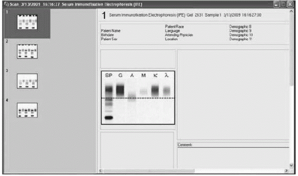
[0105] (1) Immunofixation electrophoresis assay and scanning

[0106] 1. Methodological principle: Immunofixation eletrophoresis (IFE) performs zone electrophoresis on serum or other specimens on agar (cellulose acetate) plates, and various single proteins or composite protein components are electrophoresed according to their charge, The electrical mobility is different due to the molecular size and so on. Then add the known corresponding monovalent antiserum (containing anti-κ, λ light chain and various heavy chain antiserum respectively) on it. When the antibody binds to the monoclonal Ig in its zone, an antigen-antibody complex is formed and precipitated. (fixed) down. After rinsing and staining, a thick and narrow coloring zone appears, and the light chain and heavy chain type of the monoclonal Ig can be judged.

[0107] 2. Specimen collection: 35 cases of M-proteinemia samples were collected and 1...

Embodiment 2

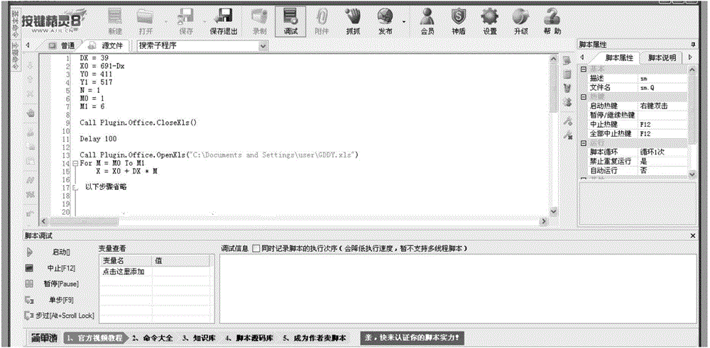
[0123] Example 2 Identification of immunofixation electrophoresis images by computer

[0124] The authors and clinical immunologists selected 35 cases of M-proteinemia sample collection and 15 cases of immunofixation electrophoresis for publication (see literature: Song Jianqing. Clinical application of electrophoresis technology—electrophoretic atlas [M]. Version 1. Liaoning: Science Press, 2006) were provided by the Laboratory of the First People's Hospital Affiliated to Shanghai Jiao Tong University. The selection principle is not only satisfied with the M protein types of routine inspection, but also including rare types and special images on the atlas of the book. After scanning into pictures, the pictures are converted into data with the button wizard computer software. Intelligent identification of immunofixation electrophoresis M protein components by computer programming Through the data analysis of 50 immunofixation electrophoresis images and serum protein electropho...


PUM


Abstract

The invention belongs to the field of clinical differential diagnosis and particularly relates to a method and a system for identifying immunofixation electrophoresis M protein components by using a computer. A sample picture subjected to immunofixation electrophoresis is collected and then is scanned by the computer to be converted into six peak-shaped electrophoretograms; the six electrophoretograms are combined by using different colors to form one electrophoretogram; an M protein zone is found firstly, and then comprehensive matching treatment is carried out according to image information of other five different components. Automatic identification, classification and comprehensive analysis of serum protein electrophoresis and all components in the immunofixation electrophoretograms can be realized by using computer programming to obtain an accurate identification result. The clinical application accuracy of the method for identifying the immunofixation electrophoresis M protein components by using the computer is 96%; compared with a traditional method, the method has no remarkable difference in the aspect of statistics; the subjective errors caused by the fact that the current traditional method is used for judging the immunofixation electrophoresis M protein components by naked eyes are greatly avoided, and extreme convenience is provided for a clinical laboratory.

Description

technical field [0001] The invention relates to a method and a system for identifying components of immunofixed electrophoresis M protein by using a computer, and specifically belongs to the field of clinical differential diagnosis. Background of the invention [0002] With the continuous development of computer technology, there are more and more applications of computer technology in medicine. Among them, the detection of automatic identification of M protein components is an important part of medical testing. The diagnosis and treatment of M proteinemia mainly depends on laboratory work. Personnel identify and classify based on zone electrophoresis and visual inspection of immunofixation electrophoresis patterns. This kind of work is inefficient and has many subjective factors, which can easily cause wrong identification. At present, domestic and foreign scholars have seldom studied this aspect. They only conduct extensive research on the bands of the Western blotting m...

Claims


Application Information

Patent Timeline
IPC IPC(8): G06F19/10
Inventor 谢松业谢婧刘庆中栾超琦黄盛葛永新陆元善俞华丁星陈频
Owner SHANGHAI FIRST PEOPLES HOSPITAL
Why Eureka
Try Eureka