X-ray diagnostic device and arm control method
A diagnostic device and a control method technology, which is applied in the direction of radiological diagnostic equipment control, radiation diagnostic diaphragm, radiation beam guiding device, etc., and can solve the problems of reduced visual recognition
- Summary
- Abstract
- Description
- Claims
- Application Information
AI Technical Summary
Problems solved by technology
Method used
Image
Examples
no. 1 Embodiment approach
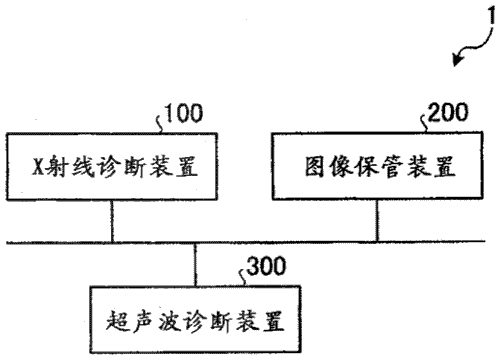
[0027] Hereinafter, details of the image processing device according to the present invention will be described. In addition, in the first embodiment, a system including the X-ray diagnostic apparatus according to the present invention will be described as an example. figure 1 It is a figure which shows an example of the structure of the system concerning 1st Embodiment.
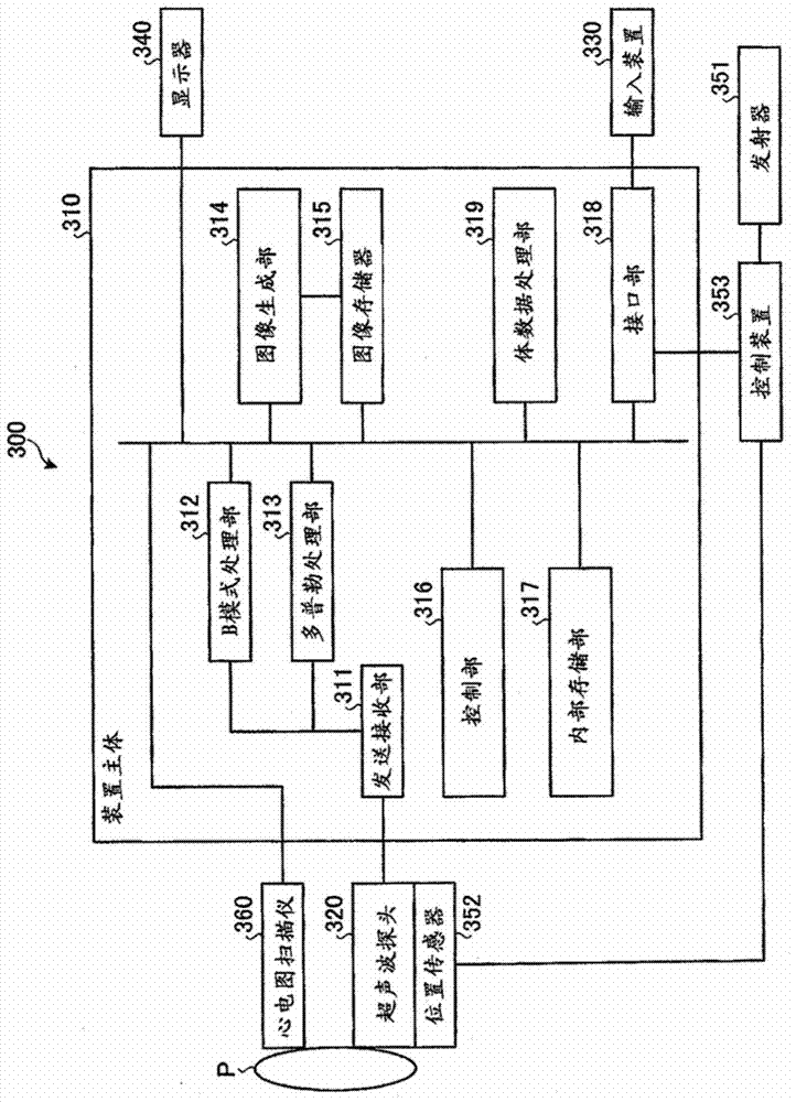
[0028] Such as figure 1 As shown, the system 1 according to the first embodiment includes an X-ray diagnostic device 100 , an image storage device 200 , and an ultrasonic diagnostic device 300 . figure 1 Each of the illustrated devices can communicate with each other directly or indirectly through, for example, a LAN (Local Area Network) installed in a hospital. For example, when a PACS (Picture Archiving and Communication System) is introduced into the image processing system 1, each device transmits and receives medical images and the like to each other in accordance with the DICOM (Digital Imaging and C...
no. 2 Embodiment approach
[0103] In the first embodiment described above, the case where the intersection point of the centers of a plurality of X-ray images is used as the isocenter has been described. In the second embodiment, a case where the position of the isocenter is moved to the center of the heart will be described. That is, in the X-ray diagnostic apparatus 100 according to the second embodiment, the coordinates of the isocenter are extracted from a plurality of X-ray images, and the C-arm or the bed is moved so that the center of the heart is located at the extracted coordinates.
[0104] The calculation unit 121b according to the second embodiment calculates the positional relationship between the center of the heart and the isocenter included in the ultrasonic image. Specifically, the calculation unit 121b acquires the coordinates of the center of the heart in the ultrasound coordinate system based on the information of the ventricles acquired by the ultrasound diagnostic apparatus 300 . ...
no. 3 Embodiment approach
[0113] In the first and second embodiments described above, the case of calculating the angle formed by the treatment site and the isocenter has been described. In the third embodiment, the case of calculating the angle formed by the treatment site and the center of the heart will be described.
[0114] The calculation unit 121b according to the third embodiment calculates the positions of the treatment site and the center of the heart in the X-ray coordinate system. Specifically, the calculation unit 121b calculates the coordinates of the treatment site in the X-ray coordinate system from the X-ray image and the ultrasonic image aligned by the alignment unit 121a. In addition, the calculation unit 121b acquires the coordinates of the heart center in the ultrasound coordinate system, and calculates the coordinates of the heart center in the X-ray coordinate system. Then, the calculating unit 121b calculates a straight line connecting the calculated coordinates of the treatmen...
PUM
 Abstract
Description
Claims
Application Information
 - R&D
- Intellectual Property
- Life Sciences
- Materials
- Tech Scout
- Unparalleled Data Quality
- Higher Quality Content
- 60% Fewer Hallucinations
Browse by: Latest US Patents, China's latest patents, Technical Efficacy Thesaurus, Application Domain, Technology Topic, Popular Technical Reports.
© 2025 PatSnap. All rights reserved.Legal|Privacy policy|Modern Slavery Act Transparency Statement|Sitemap|About US| Contact US: help@patsnap.com


