A method for visual reconstruction of the cruciate ligament of the knee joint

A cruciate ligament and knee joint technology, applied in the medical field, can solve problems such as complex operations, poor accuracy, and low efficiency, and achieve the effect of improving surgical results and avoiding measurement deviations

Active Publication Date: 2017-02-01
WEST CHINA HOSPITAL SICHUAN UNIV
View PDF6 Cites 0 Cited by
  • Summary
  • Abstract
  • Description
  • Claims
  • Application Information

AI Technical Summary

Problems solved by technology

These methods have two major disadvantages: first, it is not a digital fusion image in the true sense, with strong subjectivity and poor accuracy, to a certain extent, it is only equivalent to "paper-cutting"; second, the operation is complicated and inefficient, and batch processing cannot be realized deal with

Method used


Image

  • A method for visual reconstruction of the cruciate ligament of the knee joint

Examples


Embodiment Construction

[0023] In order to make the object, technical solution and advantages of the present invention clearer, the present invention will be further described in detail below in conjunction with the accompanying drawings.

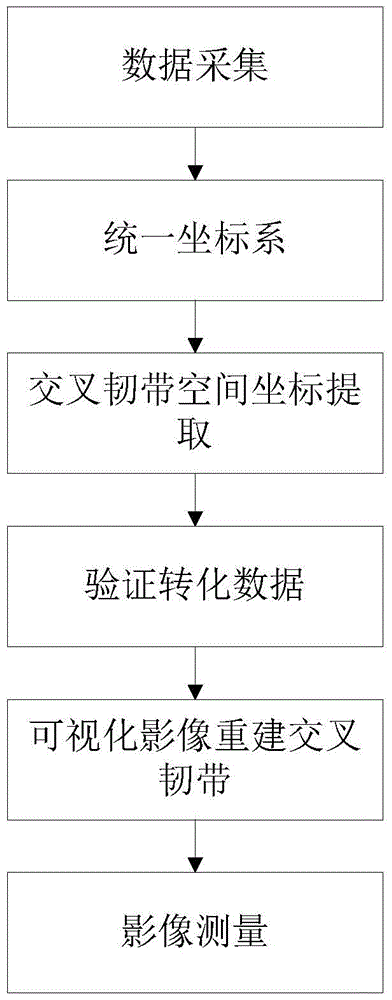
[0024] Such as figure 1 As shown, the method for visually reconstructing the cruciate ligament of the knee joint disclosed by the present invention, the specific implementation is carried out according to the following steps:

[0025] 1. Data acquisition: Use 64-slice spiral CT machine and 1.5T superconducting magnetic resonance to obtain original imaging data, fix the target knee joint at 0° straight (straight plastic plate can be used to fix it), at 30 CT and MRI scans were performed consecutively within minutes. MRI uses special limb coils, and selects T2 phase three-dimensional fast spin echo sequence (sampling perfection with application-optimized contrasts by using different flip angle evolutions, SPACE) for sagittal scanning. Scanning parameters: TR 1300m...


PUM


Abstract

The invention discloses a visual knee joint cruciate ligament reconstruction method which comprises the steps of data acquisition, coordinate system unification, cruciate ligament spatial coordinate extraction, visual cruciate ligament reconstruction based on images, image measurement and the like. The method is capable of realizing CT (Computed Tomography) and MRI (Magnetic Resonance Imaging) digital fusion, is simple and effective, and is capable of guiding knee join ligament surgery without increasing the burden of patients.

Description

technical field [0001] The invention relates to the medical field, in particular to a method for visually reconstructing the cruciate ligament of the knee joint. Background technique [0002] An ideal reconstruction of the cruciate ligament of the knee should fix the graft at its anatomical insertion point and simulate its normal shape, which requires a comprehensive understanding of the patient's individual anatomical data, especially the condition of the target ligament and its adjacent bone structure. Among various imaging techniques, computed tomography (CT) is better at visualizing bone structures, while magnetic resonance imaging (MRI) has higher resolution of soft tissues. Due to the different imaging principles of MRI and CT, it is difficult to achieve integration, so there is no CT and MRI all-in-one machine in the world. In clinical practice, when both CT and MRI are needed, the two can only be put together and interpreted by medical staff based on experience, whi...

Claims


Application Information

Patent Timeline
Patent Type & Authority Patents(China)
IPC IPC(8): A61F2/08
Inventor 陈刚李箭宁净张慧滔唐新李棋
Owner WEST CHINA HOSPITAL SICHUAN UNIV
Why Eureka
Try Eureka