
A method for detecting the laterally restricted area of ​​membrane proteins in living plants

A technology of laterally restricted area and cell membrane, which is applied in the field of detection of laterally restricted area of ​​living plant cell membrane proteins, can solve the problems that have not been clearly reported, and achieve the effect of simple method and good repeatability

Inactive Publication Date: 2017-11-28
INST OF BOTANY CHINESE ACAD OF SCI
View PDF0 Cites 0 Cited by
  • Summary
  • Abstract
  • Description
  • Claims
  • Application Information

AI Technical Summary

Problems solved by technology

However, how to achieve this purpose has not been clearly reported in the prior art

Method used


Image

  • A method for detecting the laterally restricted area of ​​membrane proteins in living plants
  • A method for detecting the laterally restricted area of ​​membrane proteins in living plants
  • A method for detecting the laterally restricted area of ​​membrane proteins in living plants

Examples


Embodiment 1

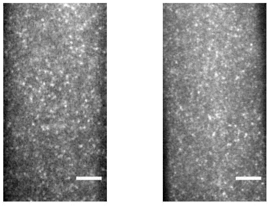
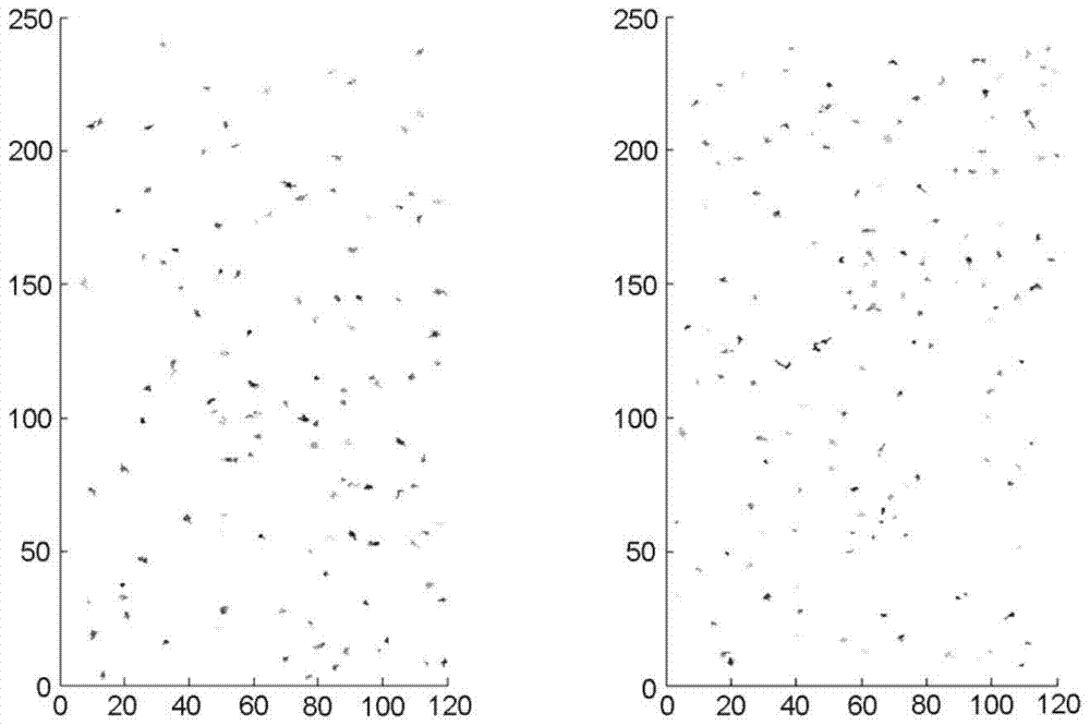
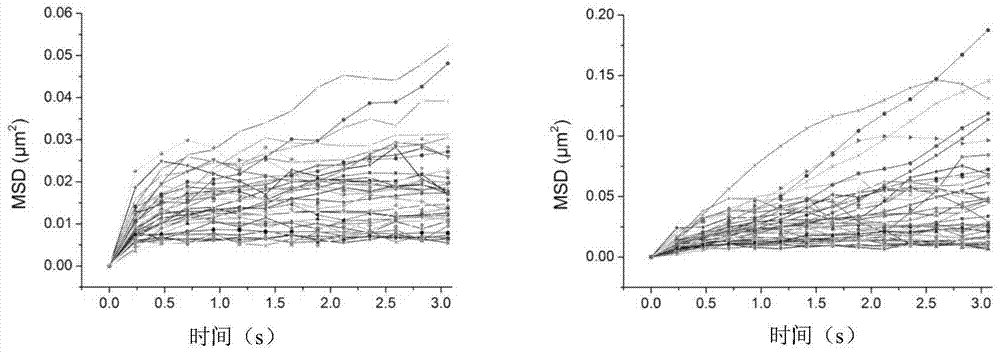
[0038] Example 1. Detection of the laterally restricted area of ​​phototropin phot1 on the cell membrane in Arabidopsis

[0039] The protein used to detect the laterally restricted area on the membrane is the Arabidopsis phototropin phot1, which is expressed on the epidermal cell membrane of the Arabidopsis hypocotyl. The phot1 tag was tagged using the green fluorescent protein GFP.

[0040] 1. Plant material

[0041] 1. Obtaining transgenic Arabidopsis

[0042] The Arabidopsis thaliana transformed into the fusion gene phot1-GFP in the background of the phot1-5 mutant used in the present invention is disclosed in the document "Sakamoto and Briggs. Cellular and subcellular localization of phototropin1. Plant Cell, 2002, 14:1723-1735" However, the public can obtain it from the Institute of Botany, Chinese Academy of Sciences.

[0043] 2. Cultivation of Arabidopsis seeds

[0044] Transgenic Arabidopsis seeds were placed in a 1.5ml centrifuge tube, soaked in 2.5% (volume fract...


PUM


Abstract

The invention discloses a method for detecting the laterally limited area of ​​a living plant cell membrane protein. The method comprises: (1) expressing a fusion protein in a plant to obtain a transgenic plant material with single-color fluorescent labeling for the protein to be tested; (2) observing the transgenic plant material with a variable-angle total internal reflection fluorescence microscope, Obtain time series images; (3) use single particle tracking technology to identify and track fluorescent points in time series images, obtain the trajectory of fluorescent points over time, and then calculate the average square displacement of fluorescent points over time; (4) use The Origin software fits the average square displacement of the fluorescent point over time, obtains the corresponding constant, and calculates the moving range of the fluorescent point. The method for calculating the laterally restricted area of ​​membrane proteins in living cells is simple and repeatable, and individual information of proteins and group average information can also be calculated.

Description

technical field [0001] The invention belongs to the field of biotechnology, and relates to a method for detecting the lateral confinement area of ​​living plant cell membrane proteins. Background technique [0002] Many important functions of the plasma membrane are closely related to the fluidity of the membrane. As an important part of the plasma membrane, membrane proteins can undergo lateral diffusion and endocytosis cycles, and play a key role in signal transmission and material transport through these movements. Therefore, detecting the distribution and diffusion movement of membrane proteins has become an important content in the study of biofilms. However, this rapid microsecond-scale motion on the nanoscale membrane structure cannot be observed with conventional microscope imaging. [0003] Through fluorescent labeling, the use of high-resolution single-molecule imaging technology can realize in vivo imaging of the dynamics of membrane protein molecules at the nan...

Claims


Application Information

Patent Timeline
Patent Type & Authority Patents(China)
IPC IPC(8): C12Q1/02C12N15/82
Inventor 林金星薛轶群王晓华张亮
Owner INST OF BOTANY CHINESE ACAD OF SCI
Learn More