Dual-imaging magnetic tweezer system

A magnetic tweezers, imaging technology, applied in material excitation analysis, fluorescence/phosphorescence, instruments, etc., can solve problems such as detection of fluorescent signal of sample molecules.

Inactive Publication Date: 2015-10-28
HUAZHONG UNIV OF SCI & TECH
View PDF11 Cites 6 Cited by
  • Summary
  • Abstract
  • Description
  • Claims
  • Application Information

AI Technical Summary

Problems solved by technology

[0005] Aiming at the problems existing in the prior art, the embodiment of the present invention provides a dual-imaging magnetic tweezers system, which is used to solve the problem that in the prior art, the sample molecules cannot be detected by fluorescent signals, but the sample molecules can also be mechanically manipulated. question

Method used


Image

  • Dual-imaging magnetic tweezer system
  • Dual-imaging magnetic tweezer system
  • Dual-imaging magnetic tweezer system

Examples


Embodiment 1

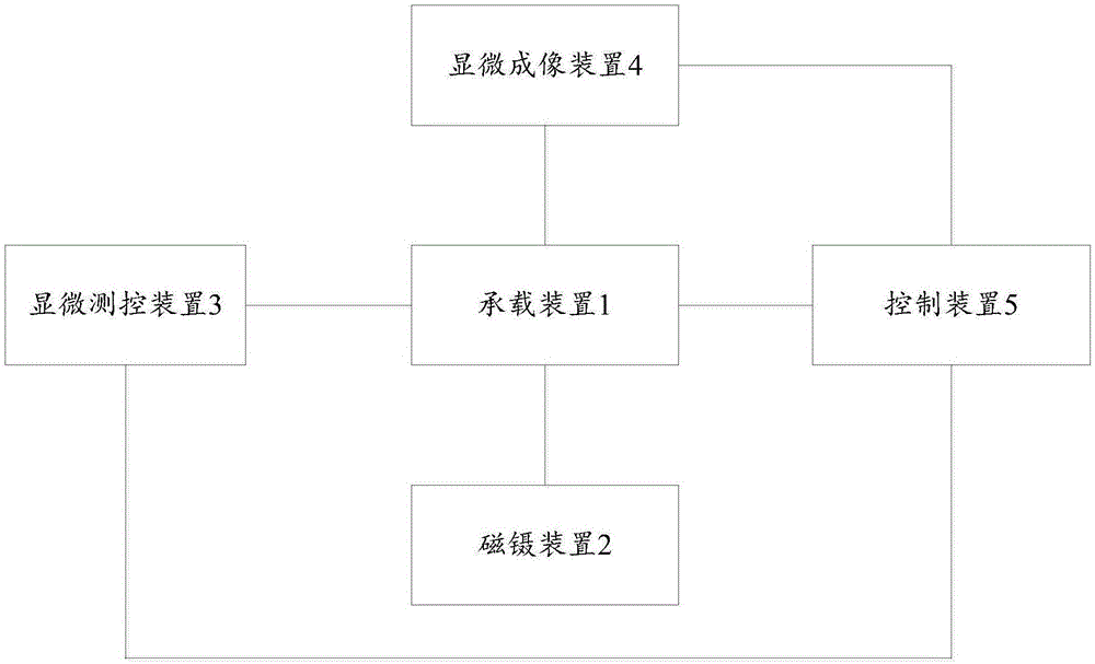
[0047] This embodiment provides a dual imaging magnetic tweezers system, such as figure 1 As shown, the system includes: a carrying device 1, a magnetic tweezers device 2, a microscopic measurement and control device 3, a microscopic imaging device 4 and a control device 5; wherein,

[0048] The carrying device 1 is used to carry sample molecules so that the system can test the sample molecules.

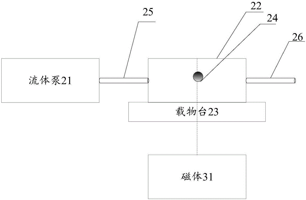
[0049] Specifically, such as figure 2 As shown, the carrying device 1 includes: a fluid pump 21, a sample tank 22, a stage 23 and a magnetic ball 24; wherein,

[0050] When the sample molecule needs to be tested, the other end of the sample molecule is connected to the magnetic ball 24 in the buffer solution, and the buffer solution is connected to the sampling tube 25 of the sample tank 22, through the The fluid pump 21 extracts the sample molecules so that the sample molecules enter the sample tank 22 along the sampling tube 25; when the sample molecules fill the sample tank 22,...

Embodiment 2

[0075] In practical applications, when the sample molecule is a λ-DNA molecule, one end of the λ-DNA is labeled with biotin, and can be connected to a magnetic ball 24 coated with streptavidin protein on the surface; the λ-DNA The other end of the DNA is marked with a sulfhydryl group and can be connected to the surface of the sample tank 22 . The testing process for the λ-DNA molecule is as follows:

[0076] First, the other end of the λ-DNA molecule is connected to the magnetic ball 24 in the buffer solution, the buffer solution is connected to the sampling tube 25 of the sample tank 22, and the fluid pump 21 is used to The λ-DNA molecule is extracted so that the λ-DNA molecule enters the sample tank 22 along the sampling tube 25; when the λ-DNA molecule fills the sample tank 22, the fluid pump 21 is closed, and the sample tank 22 Invert for a while, so that one end of the λ-DNA molecule is connected to the upper surface of the sample chamber 22 . The diameter of the magne...

Embodiment 3

[0084] Compared with Embodiment 2, the sample molecules in this embodiment are vascular endothelial cells. One end of the vascular endothelial cells can be connected to the magnetic ball 24 coated with fibronectin, and the other end can be connected to the surface of the sample chamber 22 . The testing process for the vascular endothelial cells is as follows:

[0085]First, the other end of the vascular endothelial cells is connected to the magnetic ball 24 in a buffer, the buffer is connected to the sampling tube 25 of the sample tank 22, and the fluid pump 21 is used to The vascular endothelial cells are extracted so that the vascular endothelial cells enter the sample tank 22 along the sampling tube 25; when the vascular endothelial cells fill the sample tank 22, the fluid pump 21 is turned off, and the sample tank 22 is inverted for a period of time, One end of the vascular endothelial cells is connected to the upper surface of the sample well 22 . The diameter of the mag...


PUM


Abstract

The invention provides a dual-imaging magnetic tweezer system. The dual-imaging magnetic tweezer system comprises a bearing device, a magnetic tweezer device, a microscopic measuring and controlling device, a microscopic imaging device and a control device, wherein the bearing device bears sample molecules; the magnetic tweezer device performs mechanical manipulation on the sample molecules to enable the sample molecules to move; the microscopic measuring and controlling device collects first image data of the movement of the sample molecules from the side faces of the sample molecules; the microscopic imaging device collects second image data of the movement of the sample molecules from the positions above the sample molecules; the control device analyzes the first image data to obtain a first analysis result, analyzes the second image data to obtain a second analysis result and verifies the first analysis result and the second analysis result to determine a result of the sample molecules changing with time in the three-dimensional space. Accordingly, fluorescence signal detection can be performed on the sample molecules through the microscopic measuring and controlling device, and meanwhile the mechanical manipulation can be performed on the sample molecules through the microscopic imaging device; in addition, verification can be performed according to the obtained first image data and the obtained second image data, and the test precision is improved.

Description

technical field [0001] The invention belongs to the technical field of microscopic detection, in particular to a dual-imaging magnetic tweezers system. Background technique [0002] In the study of the dynamics of biomolecules and cells at the micro-nano scale, the study of molecular conformation and conformational changes over time can further reveal the mysteries of life activities. [0003] In recent years, with the continuous development of single-molecule manipulation and detection technology, magnetic tweezers is a widely used single-molecule manipulation device. It fixes the two ends of the experimental molecule on a glass slide and a magnetic bead respectively. An external magnetic field is then applied to control the magnetic balls, stretching or twisting the experimental molecules. Through the change of the position of the magnetic ball, the length, force and torque of the experimental molecule can be calculated. Although the existing technology can perform fluor...

Claims


Application Information

Patent Timeline
Patent Type & Authority Applications(China)
IPC IPC(8): G01N21/64G01N21/01
Inventor 肖波涛白慧玲符青山
Owner HUAZHONG UNIV OF SCI & TECH
Why Eureka
Try Eureka