X-ray tomographic scanner

An X-ray and scanner technology, applied in the field of X-ray tomography scanners, can solve the problems of CT image artifacts, increased X-ray radiation dose, and high radiation dose of radiation, achieve high utilization rate, accelerate acquisition speed, and avoid radiation radiation Effect

Inactive Publication Date: 2015-11-11
TIANJIN FUSITE TECH
View PDF0 Cites 4 Cited by
  • Summary
  • Abstract
  • Description
  • Claims
  • Application Information

AI Technical Summary

Problems solved by technology

In the process of CT scanning, when data acquisition is required, the patient is absolutely still, but in fact, the patient will have voluntary or involuntary movement during the scan, which will destroy the consistency and integrity of the projection data. Once the data acquisition process Motion in the center will cause serious artifacts on the final CT image, and even affect the doctor's diagnosis
This problem is more likely to occur in cone-beam CT with longer data acquisition time
Usually, because the operator and the scanning patient are not in the same room, it is difficult to detect the motion in time during the scanning process. Often, it is judged whether t...

Method used


Image

  • X-ray tomographic scanner
  • X-ray tomographic scanner

Examples


Embodiment Construction

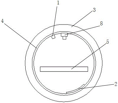
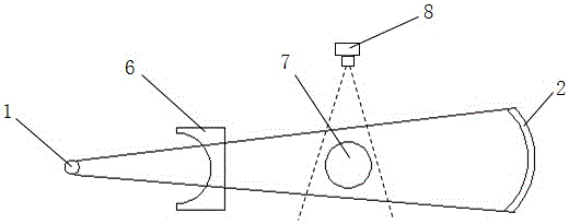
[0016] like figure 1 and figure 2 Shown, the X-ray tomography scanner of the present invention of the present invention comprises ray source 1, flat panel detector 2, detection bed 5 and collection computer, and wherein ray source emits X-ray, and flat panel detector receives X-ray and converts it into The electric signal, the flat-panel detector is connected with the acquisition computer, the test bed supports the patient 7, the fixed support 3 is arranged around the test bed, and the contact slip ring 4 is arranged inside the fixed support, and the radiation source and the flat-panel detector are respectively fixed on the contact slip ring , and the radiation source and the flat panel detector are set opposite to each other with the same diameter and rotate synchronously, and the contact slide rotates around the detection bed; a motion detection device 8 is arranged directly above the fixed bracket.

[0017] The ray source is a pulsed, rotating anode X-ray source that emit...


PUM


Abstract

An X-ray tomographic scanner comprises a ray source, a flat panel detector, a detection bed and a collection computer, wherein the ray source emits X-rays, the flat panel detector receives the X-rays, converts the X-rays into electric signals and is connected with the collection computer, and the detection bed supports a patient; a fixed support is arranged around the detection bed, a contact slip ring is arranged at the inner side of the fixed support, the ray source and the flat panel detector are fixedly arranged on the contact slip ring and arranged opposite with the same diameter and rotate synchronously, the contact slip ring rotates around the detection bed, and a movement detection device is arranged over the fixed support. Images collected by the ray source and the flat panel detector during rotation are two-dimensional projection, and the two-dimensional projection is reconstructed in a three-dimensional mode through the collecting computer to obtain three-dimensional images used for diagnosis of doctors. The use ratio of the X-rays is higher, the speed of collecting data is higher, and movement of the patient can be monitored at all times through the movement detection device.

Description

technical field [0001] The invention relates to the technical field of clinical medical equipment, in particular to a movable X-ray tomographic scanner with low radiation dose. Background technique [0002] Computed tomography (CT) is an important non-destructive testing technology, which can obtain two-dimensional or three-dimensional images of the internal and external structural features of objects by using X-ray projection data from multiple projection angles. With the advantages of non-destructive, high-precision, and three-dimensional visualization, it has been widely used in clinical medical imaging diagnosis, and the image provides an important basis for the diagnosis of patients' diseases. But now many hospitals need to make an appointment for CT examination, which cannot meet the needs of severe and acute patients, and is not conducive to the timely treatment of acute patients; as my country gradually enters an aging society, hospital resources will be more scarce ...

Claims


Application Information

Patent Timeline
IPC IPC(8): A61B6/03
Inventor 张励曹文田
Owner TIANJIN FUSITE TECH
Why Eureka
Try Eureka