[0024] The technical solutions of the embodiments of the present invention will be clearly and completely described below in conjunction with the accompanying drawings. Obviously, the described embodiments are only a part of the embodiments of the present invention, rather than all the embodiments.
[0025] According to an embodiment of the present invention, a method for detecting EGFRvIII in tumor tissue includes the following steps:
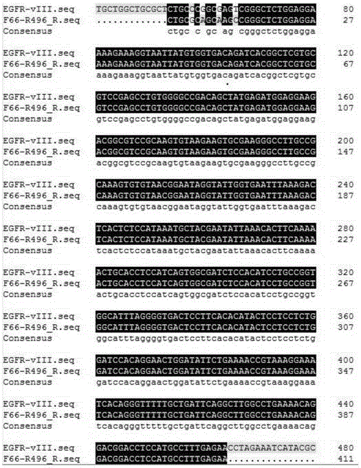
[0026] 1. EGFRvIII gene sequence analysis and primer design
[0027] Use the written sequence analysis program to scan the gene sequence before the mutation region (GC content 71.6%, high repetition rate) base by base to obtain the 56-70 base low repetitive region, use sequence analysis software to design specific reaction The primers are matched with the forward primers F56 (SEQIDNO:1) and F66 (SEQIDNO:3) before the mutation region. The NCBI primer search software is used to detect the specificity of each paired primer in the human genome. The first and second rounds of specific reverse amplification primers R796 (SEQ ID NO: 2) and R496 (SEQ ID NO: 4) of the mutant region;
[0028] Table 1: PCR detection primers
[0029]
[0030] 2. Acquisition of detection template
[0031] It has been reported that PCR methods for detecting EGFR and its mutants mostly use DNA for direct amplification. Because it contains many introns, both specificity and accuracy will be greatly compromised. Moreover, the genomic level cannot reflect the expression of EGFRvIII. Therefore, we have adopted the detection of EGFRvIII at the expression level to detect the expression of EGFRvIII in tumor tissues more intuitively and accurately, laying the foundation for targeted therapy of malignant tumors.
[0032] 1) The extraction of RNA from tissue samples includes the following steps:
[0033] 1.1) Selection and preparation of tissue samples: surgically cut tumor tissue, excise the edges, select the intact tissue block in the middle, and place it in RNAlater in the shortest time and store it at -80°C to prevent RNA degradation;
[0034] 1.2) Take 30mg of RNAlater to save the sample, add 1ml of TRIzol to fully grind it to fully lyse the tissue block, add chloroform at the ratio of 200μl/ml of TRIzol, shake and mix thoroughly, and centrifuge at 4°C, 12000rpm for 15 minutes;
[0035] 1.3) Take the upper water phase into another EP (centrifuge) tube, add an equal volume of isopropanol to precipitate RNA, centrifuge at 4°C, 12000rpm for 10 minutes, and collect the precipitated RNA;
[0036] 1.4) Wash the precipitated RNA with 75% ethanol and centrifuge at 12000rpm for 10 minutes at 4°C;
[0037] 1.5) Discard the ethanol, and when the ethanol is exhausted, dissolve the RNA of the tissue sample with an appropriate amount of DEPC water;
[0038] 2) Obtaining template cDNA, including:
[0039] 2.1) Accurately measure the RNA concentration of tissue samples;
[0040] 2.2) The instructions for the high-capacity cDNA reverse transcription kit are strictly equipped with the reverse system on ice;
[0041] Table 2: cDNA acquisition reaction system
[0042]
[0043] Gently mix the above reaction system, and centrifuge briefly in a centrifuge to obtain template cDNA;
[0044] Table 3: cDNA acquisition reaction program
[0045]
[0046] 2.3) Configure the reaction system with Taq high-fidelity DNA polymerase, the primers are F (SEQIDNO: 5) and R (SEQ ID NO: 6), and the human internal reference gene (GAPDH) is used for PCR amplification of each tissue sample, and the product is detected on a 1% gel. To ensure the quality of RNA extraction and the quality of cDNA obtained;
[0047] Table 4: GAPDH amplification system
[0048]
[0049] Table 5: GAPDH amplification program
[0050]
[0051] 3. PCR detection of EGFRvIII
[0052] From the gene sequence analysis, we can clearly get: EGFRvIII is 801bp missing from wild-type EGFR, and the PCR products of each tissue sample can be obtained by PCR. Through gel detection, we can visually observe the mutation and rough expression of EGFRvIII.
[0053] The nested PCR method amplifies the expression signal of the gene through two rounds of PCR, and due to the particularity of primer design, fully guarantees the specificity of the amplified product to ensure high accuracy and sensitivity of PCR detection;
[0054] 3.1) Using the cDNA strand obtained in step 2 as a template, prepare the first round of reaction system referring to the GAPDH amplification system, and perform the first round of PCR amplification as follows:
[0055] Table 6: The first round of PCR amplification program
[0056]
[0057] 3.2) Take 1μl of the first round of PCR product, dilute it with sterile water 10 times, and mix it thoroughly to obtain the second round of PCR template. Refer to the GAPDH amplification system to prepare the second round of PCR amplification system and perform the second round of PCR amplification. The amplification procedure is as follows:
[0058] Table 7: The second round of PCR amplification program
[0059]
[0060] 3.3) 1% agarose gel detection of the second round of amplification results, because EGFRvIII is 801bp missing from wild-type EGFR, the expression of EGFRvIII can be visually observed from the gel photograph results.
[0061] Such as figure 1 As shown, from left to right is the DL2000 standard reference, samples No. 1-21, each sample to be tested can amplify a 582bp band;
[0062] Such as figure 2 As shown, 1% agarose gel electrophoresis was used to detect the second round of nested PCR amplification products. Among them, the samples from left to right are: Dl2000 standard reference, No. 1-21 sample, negative control sample (water) and positive Control sample; the 23rd lane is a negative control, and no band is amplified using water as a template; the 24th lane is a band with the 431bp template size amplified in the known tumor cells of the EGFRvIII gene; No.1-4 Samples, 6, 7, 11, 16, 19, 20, and 21 samples can amplify a 431bp band.
[0063] 4. Sequencing verification of test results, including steps:
[0064] 4.1) Sequencing sample acquisition: accurately cut gel to recover EGFRvIII bands and recover EGFRvIII gene fragments; accurately measure the concentration of recovered gene fragments to ensure that the concentration required for sequencing is reached;
[0065] 4.2) Sequencing to confirm PCR results;
[0066] 4.3) Perform an overall sequence analysis based on the sequencing results and EGFR and EGFRvIII sequences to determine the accuracy of PCR amplification.
[0067] Such as image 3 As shown, the second round of nested PCR products are sequenced and analyzed, and sequence analysis software is used to compare the sequencing results. Among them, F66-496 is the sequencing result sequence of the second round of nested PCR products. The ellipsis indicates that compared with the EGFRvIII gene sequence, the measured sequence is shorter than EGFRvIII because the measured sequence is part of the EGFRvIII gene sequence; The second line is the sequence of the target EGFRvIII gene, the blue part indicates that the two sequences are completely matched, the cyan part indicates that the two sequences do not match; the third line is the consensus sequence of the first two sequences; the front end is the sequence across the mutation region (88 -89 bases). The results showed that the sequenced sequence is exactly the same as the target gene sequence, that is, the nested PCR product is a section of the EGFRvIII gene, indicating that the primer has good specificity, and the PCR amplified product is the EGFRvIII sequence.