Ultrasound systems and methods for automated fetal heartbeat identification

An ultrasonic image and imaging system technology, applied in the direction of ultrasonic/sonic/infrasonic Permian technology, organ movement/change detection, ultrasonic/sonic/infrasonic image/data processing, etc., can solve the problem of no anatomical structure, difficult to distinguish , Limiting the accuracy of fetal heart cycle measurement and other issues

Active Publication Date: 2016-05-18
KONINKLJIJKE PHILIPS NV
View PDF11 Cites 4 Cited by
  • Summary
  • Abstract
  • Description
  • Claims
  • Application Information

AI Technical Summary

Problems solved by technology

As the fetus moves, the orientation of the desired image data relative to the probe will change, and the fetal heart may move completely out of the field of view, resulting in no desired anatomy in the acquired data set
Also,

Method used


Image

  • Ultrasound systems and methods for automated fetal heartbeat identification
  • Ultrasound systems and methods for automated fetal heartbeat identification
  • Ultrasound systems and methods for automated fetal heartbeat identification

Examples


Embodiment Construction

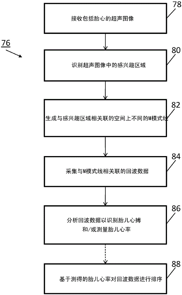
[0016] The present invention provides systems and methods for identifying heartbeats (eg, fetal heartbeats) and associated heart rates. For example, the systems and methods can be used to reduce scan time for sonographers, increase diagnostic confidence, and simplify workflow for scanning maternal patients.

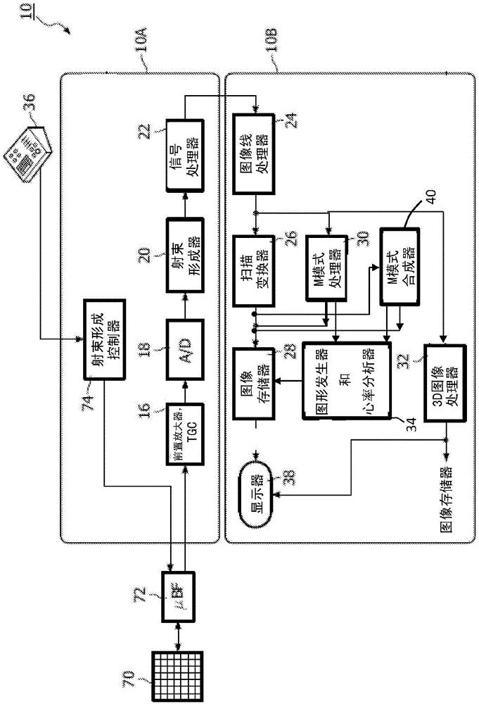
[0017] In one embodiment, the invention includes an ultrasound imaging system for identifying fetal poles or the heart and / or associated heart rate. The system of the present invention includes an ultrasound probe. A variety of probes can be used and can include array transducers. The system also includes an image processor for processing echo data from the probe. Echo data can include echo signals obtained by multiple imaging modalities, such as B-mode or M-mode image acquisition. The system is also capable of transmitting echo data and / or displaying echo data from the probe for review. An image display in the system is coupled to the image processor and adapted to d...


PUM


Abstract

Ultrasound systems and methods provide a workflow to automatically identify a fetal heartbeat. A region of interest (ROI) is identified in an ultrasound image and an ROI is identified that contains the fetal heart. The ultrasound system produces spatially different M-mode lines associated with the ROI. The ultrasound system can identify a fetal heartbeat by tracking the changing position of the heart wall and estimate the fetal heart rate, e.g. by measuring from peak-to-peak of two subsequent waves. The echo signals for the M-mode lines can also be ranked according to the likely presence of a fetal heartbeat in the echo data.

Description

technical field [0001] Ultrasound is well suited for fetal imaging because it performs non-invasive imaging without exposing the mother or fetus to ionizing radiation. The purpose of many fetal examinations is to assess the development of fetal anatomy to determine whether the fetus is developing normally. As ultrasound image quality has improved over the years, more areas of anatomy can be visualized in greater detail for developmental assessment. Accordingly, fetal ultrasonography has become more comprehensive as the demands on the anatomy to be evaluated have increased. One area of ​​the anatomy that was scrutinized was the developing fetal heart. Background technique [0002] Ultrasound systems can be used to detect fetal heartbeats up to about five weeks' gestation. At this point, the normal fetal heart rate is similar to the mother's heart rate, eg, about 80-85 beats per minute. For the next month, the heart rate will increase steadily about three times per minute ...

Claims


Application Information

Patent Timeline
IPC IPC(8): A61B8/02A61B8/08
Inventor D·A·施特拉斯纳J·曹V·T·沙姆达莎尼I·A·穆夫蒂穆罕默德
Owner KONINKLJIJKE PHILIPS NV
Why Eureka
Try Eureka