
Image omics based lesion tissue auxiliary prognosis system and method

A technology of radiomics and prediction methods, applied in computer-aided medical procedures, informatics, image analysis, etc.

Inactive Publication Date: 2016-06-08
INST OF AUTOMATION CHINESE ACAD OF SCI
View PDF7 Cites 33 Cited by
  • Summary
  • Abstract
  • Description
  • Claims
  • Application Information

AI Technical Summary

Problems solved by technology

However, there is still untapped valuable information in medical imaging to reveal lesion staging and prognosis

Method used


Image

  • Image omics based lesion tissue auxiliary prognosis system and method

Examples


Embodiment Construction

[0015] In order to make the objectives, technical solutions and advantages of the present invention more clearly understood, the present invention will be further described in detail below in conjunction with specific embodiments and with reference to the accompanying drawings.

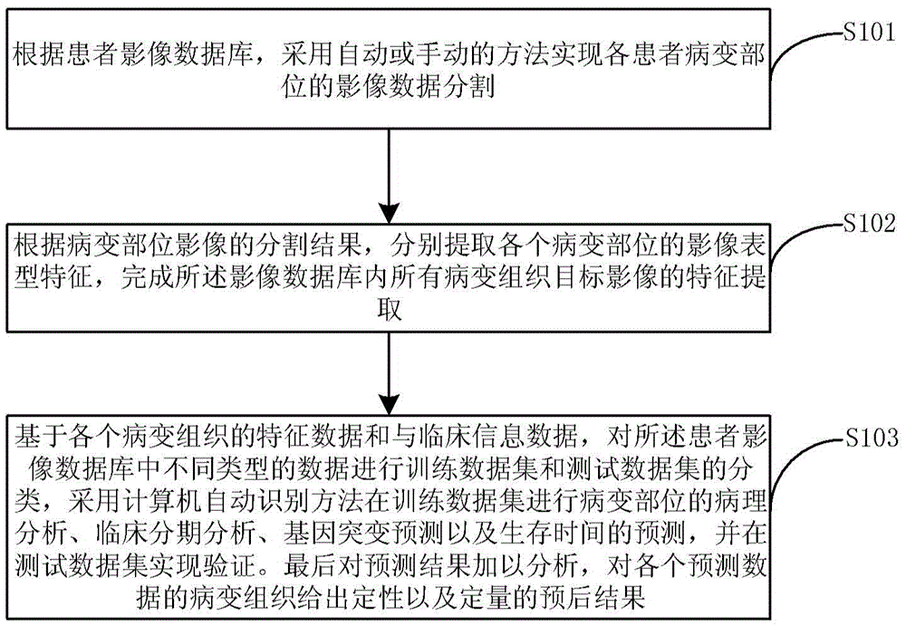
[0016] The invention discloses a method for auxiliary prognosis of diseased tissue based on radiomics. Type features, establish a complete phenotypic feature database; obtain patient tissue biopsy results, genotypes, survival time and other information according to the basic clinical information of each patient in the image database, and use computer automatic identification and classification methods to analyze the pathological manifestations, clinical features of diseased tissue. The characteristics of staging and gene mutation type are trained and classified separately to establish a reliable prediction and prognosis model; it is applied to test data and other independent data to achieve separate pr...


PUM


Abstract

The invention discloses an image omics based lesion tissue auxiliary prognosis system and method. The method comprises the steps of extracting image data of lesion parts with an automatic or manual segmentation method from a big-data-volume patient image database; according to segmentation results of lesion part images, extracting image phenotypic characteristics of each lesion part, and finishing feature extraction of image data of all lesion parts in the patient image database; and based on characteristic data and clinical information data of each lesion part, performing training data set and test data set classification on the data in the patient image database, performing pathologic analysis, clinical stage analysis, gene mutation prediction and survival time prediction of the lesion parts in the training data set with a computer automatic identification method, and performing verification in the test data set. The method is capable of performing qualitative and quantitative prediction analysis on a specific individual and providing credible prediction and analysis results.

Description

technical field [0001] The present invention relates to the technical field of disease diagnosis assistance, and more particularly to a system and method for assisting prognosis of diseased tissue based on radiomics. Background technique [0002] As a non-invasive method for early diagnosis of tumors, medical imaging has been widely used in the auxiliary diagnosis of various cancers. At present, the use of image information for clinical auxiliary diagnosis often relies on the subjective experience of the doctor, and the corresponding diagnosis is given based on the imaging characteristics of the patient's disease reflected by the image. However, there is still unexplored valuable information in medical imaging to reveal disease staging and prognosis. [0003] Different types of tumors have different manifestations on imaging due to their pathological characteristics, and different tumor imaging features also indicate completely different treatment methods and directly affec...

Claims


Application Information

Patent Timeline
Patent Type & Authority Applications(China)
IPC IPC(8): G06F19/00G06T7/00
CPCG06F19/34G06F19/321
Inventor 田捷宋江典董迪臧亚丽刘振宇
Owner INST OF AUTOMATION CHINESE ACAD OF SCI
Learn More