Helical tomotherapy image quality improvement method

A technology of image quality and tomography, applied in the field of medical image processing, can solve the problems of low contrast, high noise and unclear edges of helical tomographic radiotherapy images

Active Publication Date: 2016-06-22
BEIHANG UNIV
View PDF3 Cites 2 Cited by
  • Summary
  • Abstract
  • Description
  • Claims
  • Application Information

AI Technical Summary

Problems solved by technology

[0004] 1. Purpose: The purpose of the present invention is to provide a method for enhancing the image quality of helical tomotherapy based on the retina-cerebral cortex theory. The image quality is improved to solve the problems of more noise, low contrast and unclear edges in helical tomotherapy images

Method used


Image

  • Helical tomotherapy image quality improvement method
  • Helical tomotherapy image quality improvement method
  • Helical tomotherapy image quality improvement method

Examples


Embodiment Construction

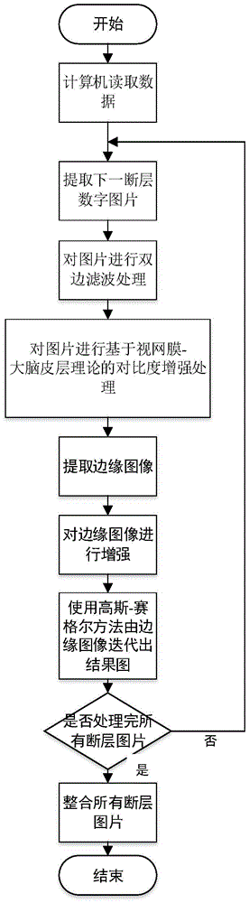
[0031] A method for improving the image quality of spiral tomotherapy based on the retina-cerebral cortex theory of the present invention, see figure 1 As shown, the steps are as follows:

[0032] Step 1: first output computer digital images through the helical tomotherapy apparatus, then use the imread function in Matlab language to read the image, and convert its information into Matlab matrix form, so that Matlab language can process it.

[0033] The helical tomotherapy image in the present invention is a digital image of 512 pixels*512 pixels*c channel, that is, the read-in matrix data has dimensions of 512*512*c. The symbol c represents the number of slices contained in the image, each slice is a grayscale image, and the slice images with the number of c channels are stacked to form a complete helical tomotherapy image. This method does not use the correlation information between channels, so the following steps are all completed on a single tomographic image. For conv...


PUM


Abstract

A helical tomotherapy image quality improvement method relates to the helical tomotherapy image quality improvement method based on the retina-cerebral cortex theory. The method mainly comprises four steps of 1 using a computer to read a helical tomotherapy image under a MATLAB environment; 2 carrying out the bilateral filtering denoising on the image; 3 carrying out the contrast promotion on the image based on the retina-cerebral cortex theory; 4 using a Gauss-Zagel iteration method to enhance the edges of the image. The helical tomotherapy image quality improvement method of the present invention enables the problem that the original helical tomotherapy image is more in noise, poor in contrast and unclear in edge to be solved, obtains a better quality enhancement result, and has the wide application prospect in the helical tomotherapy field.

Description

(1) Technical field: [0001] The invention provides a method for improving the image quality of helical tomotherapy, which relates to a method for improving the quality of helical tomotherapy images based on the retina-cerebral cortex theory, and belongs to the field of medical image processing. (two) background technology: [0002] With the development of information technology and medical imaging technology, medical image processing plays an increasingly important role in medical clinical and scientific research, and strongly promotes the progress of medical scientific research and clinical treatment. Among them, the image processing technology for CT (Computed Tomography, ie computerized tomography) images plays an important role. However, research on image processing of mega-voltage computed tomography (MVCT, Mega-Voltage Computed Tomography) images for tomotherapy is still at an early stage. Helical tomotherapy is a cancer radiotherapy method based on helical tomotherap...

Claims


Application Information

Patent Timeline
Patent Type & Authority Applications(China)
IPC IPC(8): G06T5/00
CPCG06T5/003G06T2207/10081G06T2207/20028G06T2207/30004
Inventor 史振威林浩宁夏廷毅吴伟章
Owner BEIHANG UNIV
Why Eureka
Try Eureka