Pathological image cell nucleus quick location method

A pathological image and positioning method technology, applied in the field of digital image processing of cell nucleus center positioning in digital pathological images, can solve the problem that the processing speed is difficult to meet the computer-aided analysis of digital pathological full slices, and achieve easy implementation, fast execution speed, and flow clear effect

Active Publication Date: 2016-07-06
BEIHANG UNIV
View PDF5 Cites 23 Cited by
  • Summary
  • Abstract
  • Description
  • Claims
  • Application Information

AI Technical Summary

Problems solved by technology

[0005] The present invention is a fast nuclei positioning method for pathological images. Aiming at the problem that the processing speed of the traditional nuclei positioning method is difficult to meet the computer-aided analysis of digital pathological full slices, the digital pathological image can be quickly and accurately positioned without segmenting the nuclei. nuclei in

Method used


Image

  • Pathological image cell nucleus quick location method
  • Pathological image cell nucleus quick location method
  • Pathological image cell nucleus quick location method

Examples


Embodiment Construction

[0016] In order to better understand the technical solution of the present invention, the present invention will be described in detail below in conjunction with the accompanying drawings and specific embodiments.

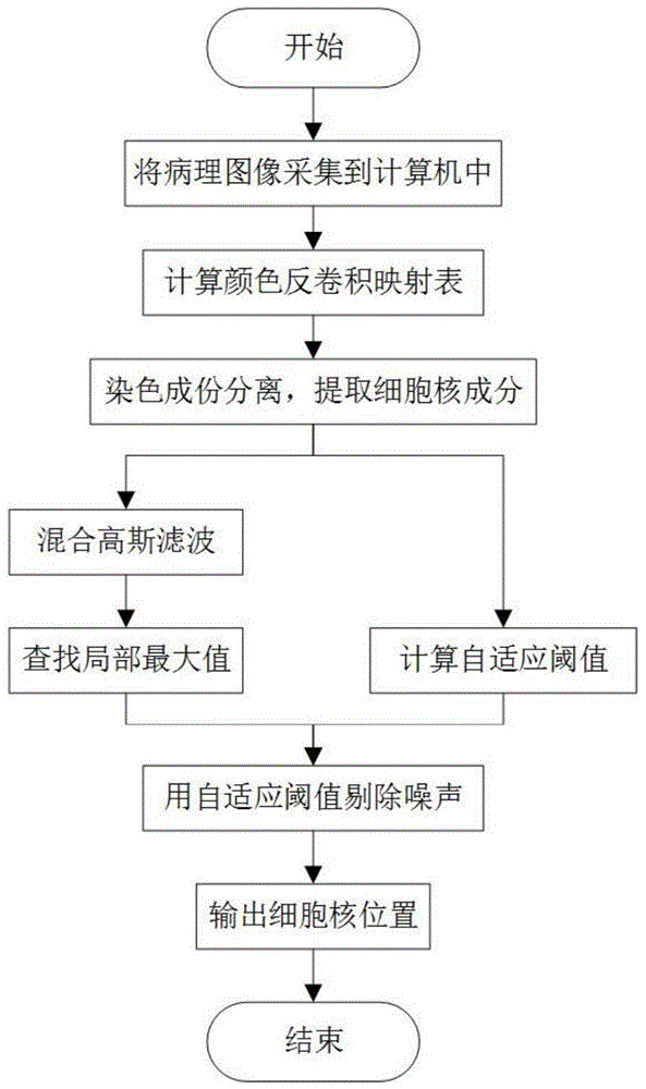
[0017] The present invention is a method for rapidly locating cell nuclei in pathological images, and the method mainly includes the following steps:

[0018] 1. Use a slice scanner to scan pathological slices for tissue biopsy into an electronic computer, and store them as a digital image matrix in the form of RGB three-channel.

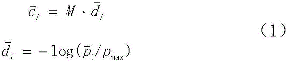
[0019] 2. Use the color deconvolution matrix to calculate the color deconvolution mapping table.

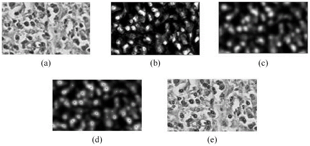
[0020] 3. Using the color deconvolution mapping table in step 2 to extract the nuclei components in the digital pathological slice, and obtain the nuclei component images.

[0021] 4. Perform mixed Gaussian filtering on the nucleus component image obtained in step 3 to obtain a filtered image.

[0022] 5. Find the local maximum in the fil...


PUM


Abstract

The invention discloses a quick and stable cell nucleus location method in order to solve the problem that the processing speed of the traditional cell nucleus location algorithm can hardly meet the requirement of large-scale digital pathological full-slice computer-aided analysis. First, a color de-convolution algorithm is used to extract the cell nucleus component in a pathological image; then, the center position of the cell nucleus is highlighted by a mixed Gaussian filter; and finally, the local maximum value is found in the filtered cell nucleus image, and the position of the cell nucleus is determined through adaptive threshold judgment. The method has the advantages of clear process, easy implementation, low time complexity, fast execution and stable location effect, and is of great significance in digital pathological full-slice computer-aided analysis.

Description

technical field [0001] The invention discloses a method for rapidly locating cell nuclei in pathological images, which belongs to the field of digital image processing, and in particular relates to a digital image processing technology for locating the center of cell nuclei in digital pathological images. Background technique [0002] Digital pathological full slide is to convert pathological slices into high-magnification large-scale digital images that can be displayed, transmitted and processed by a computer through a dedicated scanning imaging system. In cancer diagnosis based on full digital pathology slides, the morphological characteristics of cell nuclei and the distribution of different types of cell nuclei in tissues are the main basis for cancer diagnosis. Accordingly, many computer-aided cancer diagnosis methods start with the precise positioning and segmentation of cell nuclei, analyze the characteristics of tissues in digital full sections, and perform auxiliar...

Claims


Application Information

Patent Timeline
Patent Type & Authority Applications(China)
IPC IPC(8): G06T7/00
CPCG06T7/0012G06T2207/10024G06T2207/20024G06T2207/30024
Inventor 姜志国郑钰山张浩鹏麻义兵
Owner BEIHANG UNIV
Why Eureka
Try Eureka