Analyzing and processing method for analyzing and processing magnetic resonance image of acute ischemic stroke

A magnetic resonance image and ischemic stroke technology, applied in the field of image analysis and processing, can solve the problems of artifact interference, uneven distribution, blurred boundaries, etc., and achieve high segmentation accuracy, good segmentation accuracy, noise or artifacts. low impact effect

Active Publication Date: 2016-07-20
SHENZHEN INST OF ADVANCED TECH
View PDF5 Cites 8 Cited by
  • Summary
  • Abstract
  • Description
  • Claims
  • Application Information

AI Technical Summary

Problems solved by technology

In the first case, due to the influence of T2 penetration effect, some ischemic areas show abnormally high signal on DWI, which is not obvious, and the boundary is blurred and the distribution is not uniform
In the second case, the interference of artifacts
The false normalization of ADC may be manifested by the fact that the corresponding ADC value in the ischemic area does not decrease or the degree of decrease is not obvious. In addition, the area where ADC decreases does not necessarily correspond to ischemia
These will affect the segmentation accuracy of the ADC threshold segmentation method
[0006] Existing method 2: Prakash et al. use the grayscale histogram of the ischemic image to align the asymmetry of the sagittal plane, that is, the divergence (divergencemeasure, DM) to find the area where the cerebral ischemia is located, and then find the appropriate grayscale threshold to distinguish Ischemia and non-ischemia on DWI, so it is basically a global gray-level threshold segmentation method, which is difficult to adapt to the uneven gray level of DWI images in the hyperacute phase

Method used


Image

  • Analyzing and processing method for analyzing and processing magnetic resonance image of acute ischemic stroke
  • Analyzing and processing method for analyzing and processing magnetic resonance image of acute ischemic stroke
  • Analyzing and processing method for analyzing and processing magnetic resonance image of acute ischemic stroke

Examples


Embodiment Construction

[0031] In order to make the purpose, technical solutions and advantages of the embodiments of the present invention more clear, the present invention will be further described in detail below in conjunction with the embodiments and the accompanying drawings. Here, the exemplary embodiments and descriptions of the present invention are used to explain the present invention, but not to limit the present invention.

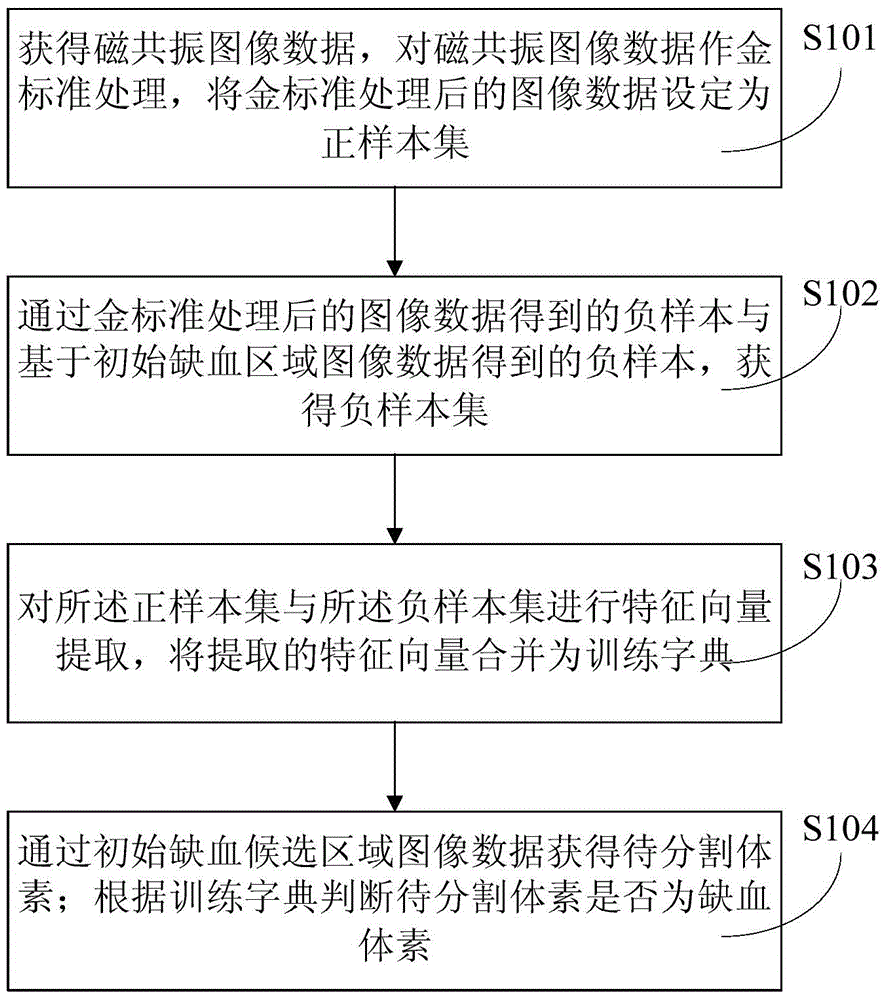
[0032] Please refer to image 3 as shown, image 3 The flow chart of the method for analyzing and processing the magnetic resonance images of acute ischemic stroke is provided for the present invention, specifically including:

[0033] S101: Obtain magnetic resonance image data, perform gold standard processing on the magnetic resonance image data, and set the image data processed by the gold standard as a positive sample set;

[0034] S102: Obtain a negative sample set through the negative sample obtained from the gold standard processed image data and the negativ...


PUM


Abstract

The invention provides an analyzing and processing method for analyzing and processing the magnetic resonance image of the acute ischemic stroke the method specifically comprises the steps of according to the data of a magnetic resonance image, acquiring the data of the magnetic resonance image, conducting the gold standard treatment on the data of the magnetic resonance image, setting the data of the image after the gold standard treatment as a positive sample set, obtaining a negative sample based on the data of the image after the gold standard treatment, obtaining a negative sample based on the data of the image in an initial ischemic region, obtaining a negative sample set, extracting the feature vectors of the positive sample set and the negative sample set, combining the extracted feature vectors as a training dictionary, obtaining to-be-segmented voxels based on the data of the image in an initial ischemic candidate region, and judging whether to-be-segmented voxels are ischemic voxels or not according to the training dictionary. According to the technical scheme of the invention, based on the analyzing and processing method for analyzing and processing the magnetic resonance image of the acute ischemic stroke, an ischemic region can be segmented more accurately. Meanwhile, the segmentation accuracy is high and the influence of noises or artifacts is small.

Description

technical field [0001] The invention relates to the field of image analysis and processing, in particular to a method for analyzing and processing magnetic resonance images of acute ischemic stroke. Background technique [0002] Ischemic stroke is the most common type of stroke. How to make a qualitative and localized diagnosis of the ischemic area of ​​ischemic stroke within the shortest time after onset, so as to guide the correct clinical diagnosis and treatment, has always been the focus of neuroimaging research. [0003] Existing MRI diffusion-weighted imaging includes: T2-weighted image (B0 image) with a diffusion coefficient of 0, and a diffusion coefficient of 1000-1500s / mm 2 The diffusion-weighted image DWI and the apparent diffusion coefficient ADC calculated by the two can reflect the imaging of the diffusion characteristics of water molecules and are very sensitive to the diffusion movement of water molecules. It is obviously better than conventional MRI in the ...

Claims


Application Information

Patent Timeline
Patent Type & Authority Applications(China)
IPC IPC(8): G06T7/00G06K9/66A61B5/055
Inventor 胡庆茂景莎莎李维平任力杰
Owner SHENZHEN INST OF ADVANCED TECH
Why Eureka
Try Eureka